
Cerebral abscess
Updates to Article Attributes
A brain abscess is a focal area of necrosis starting in an area of cerebritis surrounded by a membrane. It is a potentially life-threatening condition requiring prompt radiological identification and rapid treatment. Fortunately, MRI is usually able to convincingly make the diagnosis, distinguishing abscesses from other ring-enhancing lesions.
Epidemiology
Demographics reflect at-risk groups (see below) with all age groups being affected.
Risk factors
Risk factors for haematogenous spread include 3:
- right to left shunt
- congenital heart disease
- pulmonary AVM and AVFs as seen in hereditary haemorrhagic telangiectasia (HHT)
-
infective endocarditis
- intravenous drug use (IVDU)
- lung infection
- sinonasal infections
- dental abscess
- systemic sepsis
Clinical presentation
Clinical presentation is non-specific, with many cases having no convincing inflammatory or septic symptoms. Symptoms of raised intracranial pressure, seizures and focal neurological deficits are the most common forms of presentation. Eventually, many abscesses rupture into the ventricular system, which results in a sudden and dramatic worsening of the clinical presentation and often heralds a poor outcome.
Pathology
Cerebral abscesses result from pathogens growing within the brain parenchyma. Initial parenchymal infection is known as cerebritis, which may progress into a cerebral abscess. Historically direct extension from sinus or scalp infections was the most common source. More recently haematological spread has become most common. Direct introduction by trauma or surgery accounts for only a small minority of cases 3.
Cerebral infection is commonly divided into four stages with distinct imaging and histopathologic features:
- early cerebritis( a focal infection without a capsule or pus formation,can resolve or develop into frank abcess)
- late cerebritis
- early abscess/encapsulation - may occur 10 days after infection
- late abscess/encapsulation - may occur >14 days after infection
Microbiology
-
Streptococcus sp: 35-50% 3
- especially S. pneumoniae 4
- sterile: 25%
- mixed: variable, 10-90% of cases depending on source 3
- Staphylococcus aureus and epidermidis: following neurosurgery
- Gram-negative species more common in infants
- Listeria in pregnant women and older patients
- group B Streptococcus (GBS) and E. coli in neonates
The immunocompromised patient is susceptible to a host of other organisms including 3:
- Toxoplasma gondii
- Nocardia asteroides
- Candida albicans
- Listeria monocytogenes
- Mycobacterium spp.
- Aspergillus fumigatus
Radiographic features
Both CT and MRI demonstrate similar features, although MRI has a greater ability to distinguish a cerebral abscess from other ring-enhancing lesions:
- early cerebritis - please see cerebritis article
- late cerebritis - please see cerebritis article
-
early encapsulation
- a discrete lesion with a thin enhancing rim
- the rim may be less-well defined along the peripheral aspect of the lesion (away from ventricles)
- +/- additional "daughter" collections
- +/- ventricular extension, with accompanying ventriculitis
- late encapsulation - progressive central necrosis, cavity shrinks, decreasing surrounding oedema
CT
In patients with suspected intraparenchymal sepsis, pre- and post-contrast scans should be obtained, unless the plan is to proceed to MRI regardless of the CT findings. Typical appearances include:
- outer hypodense and inner hyperdense rim (double rim sign) in most cases
- ring of iso- or hyperdense tissue, typically of uniform thickness
- central low attenuation (fluid/pus)
- surrounding low density (vasogenic oedema)
- ventriculitis may be present, seen as enhancement of the ependyma
- obstructive hydrocephalus will commonly be seen when intraventricular spread has occurred
MRI
MRI is more sensitive than CT. Although peripherally-enhancing lesions may be non-specific by imaging, diffusion-weighted sequences (less commonly MR spectroscopy) showing central diffusion restriction are critical for suggesting the diagnosis of a cerebral abscess.
-
T1
- central low intensity (hyperintense to CSF)
- peripheral low intensity (vasogenic oedema)
- ring enhancement
- ventriculitis may be present, in which case hydrocephalus will commonly also be seen
-
T2/FLAIR
- central high intensity (hypointense to CSF, does not attenuate on FLAIR)
- peripheral high intensity (vasogenic oedema)
- the abscess capsule may be visible as an intermediate to slightly low signal thin rim 1
-
DWI/ADC
- high DWI signal is usually present centrally 11
- represents true restricted diffusion.
- peripheral or patchy restricted diffusion may also be seen; this finding is however not as constant as one may think, with up to half of the rim-enhancing lesions demonstrating some restriction not proving to be abscesses.
- in some immunocompromised states, central content may not diffusion restrict 13
-
SWI
- low-intensity rim 9
- complete in 75%
- smooth in 90%
- mostly overlaps with contrast-enhancing rim
- dual rim sign: a hyperintense line located inside the low-intensity rim
- low-intensity rim 9
- MR perfusion: rCBV is reduced in the surrounding oedema cf. to both normal white matter and tumour oedema seen in high-grade gliomas 2
- MR spectroscopy: elevated peaks are seen corresponding to lipids/lactate, succinate, acetate, and amino acids (alanine, valine, leucine, and isoleucine) 14
Treatment and prognosis
The mainstay of treatment for cerebral abscesses is neurosurgical intervention and drainage of the collection. This can be performed either by stereotactic aspiration or craniotomy 7. Broad-spectrum intravenous antibiotics are also needed and can later be changed to agents tailored to the specific organisms.
In cases where the abscess cavity does not completely obliterate, follow-up with MRI including DWI is useful and lack of restricted diffusion is reassuring. Demonstration of ongoing restricted diffusion in a cavity suggests persistent infection 8.
Differential diagnosis
The differential diagnosis typically includes other ring-enhancing lesions:
-
metastasis or high-grade glioma (e.g. GBM)
- abscesses tend to have smoother inner wall 2
- satellite lesions favour infection 2
- abscesses may have low-intensity capsule 1
-2,2 - rCBV elevated in high-grade gliomas, reduced in abscesses 2
- absent dual rim sign
- the cystic component does not show restricted diffusion, unlike abscess
- subacute infarction, subacute haemorrhage or contusion
- demyelinating lesion
- radiation necrosis
When a lesion demonstrates both ring enhancement and central restricted diffusion the differential is very much narrowed, and although cerebral abscess is by far the most likely diagnosis, the following should also be included on the differential 6:
- cerebral metastases: particularly necrotic adenocarcinoma
-<a href="/articles/pneumonia-summary">pneumonia </a><sup>12</sup>- +<a href="/articles/pneumonia-summary">pneumonia</a> <sup>12</sup>
-<li>+/- ventricular extension, with accompanying <a title="ventriculitis" href="/articles/ventriculitis">ventriculitis</a>- +<li>+/- ventricular extension, with accompanying <a href="/articles/ventriculitis">ventriculitis</a>
-<li>abscesses may have low-intensity capsule <sup>1-2</sup>- +<li>abscesses may have low-intensity capsule <sup>1,2</sup>
References changed:
- 1. Haimes A, Zimmerman R, Morgello S et al. MR Imaging of Brain Abscesses. AJR Am J Roentgenol. 1989;152(5):1073-85. <a href="https://doi.org/10.2214/ajr.152.5.1073">doi:10.2214/ajr.152.5.1073</a> - <a href="https://www.ncbi.nlm.nih.gov/pubmed/2705342">Pubmed</a>
- 2. Holmes T, Petrella J, Provenzale J. Distinction Between Cerebral Abscesses and High-Grade Neoplasms by Dynamic Susceptibility Contrast Perfusion MRI. AJR Am J Roentgenol. 2004;183(5):1247-52. <a href="https://doi.org/10.2214/ajr.183.5.1831247">doi:10.2214/ajr.183.5.1831247</a> - <a href="https://www.ncbi.nlm.nih.gov/pubmed/15505287">Pubmed</a>
- 3. Greenberg M. Handbook of Neurosurgery. (2006) ISBN: 9783131108869 - <a href="http://books.google.com/books?vid=ISBN9783131108869">Google Books</a>
- 4. Popp A, Deshaies E. A Guide to the Primary Care of Neurological Disorders. (2008) ISBN: 9781588905161 - <a href="http://books.google.com/books?vid=ISBN9781588905161">Google Books</a>
- 5. Lai P, Ho J, Chen W et al. Brain Abscess and Necrotic Brain Tumor: Discrimination with Proton MR Spectroscopy and Diffusion-Weighted Imaging. AJNR Am J Neuroradiol. 2002;23(8):1369-77. <a href="https://www.ncbi.nlm.nih.gov/pmc/articles/PMC7976258">PMC7976258</a> - <a href="https://www.ncbi.nlm.nih.gov/pubmed/12223380">Pubmed</a>
- 6. Hartmann M, Jansen O, Heiland S, Sommer C, Münkel K, Sartor K. Restricted Diffusion Within Ring Enhancement is Not Pathognomonic for Brain Abscess. AJNR Am J Neuroradiol. 2001;22(9):1738-42. <a href="https://www.ncbi.nlm.nih.gov/pmc/articles/PMC7974430">PMC7974430</a> - <a href="https://www.ncbi.nlm.nih.gov/pubmed/11673170">Pubmed</a>
- 7. Johnson R, Griffin J, McArthur J. Current Therapy in Neurologic Disease. (2006) ISBN: 9780323034326 - <a href="http://books.google.com/books?vid=ISBN9780323034326">Google Books</a>
- 8. Cartes-Zumelzu F, Stavrou I, Castillo M, Eisenhuber E, Knosp E, Thurnher M. Diffusion-Weighted Imaging in the Assessment of Brain Abscesses Therapy. AJNR Am J Neuroradiol. 2004;25(8):1310-7. <a href="https://www.ncbi.nlm.nih.gov/pmc/articles/PMC7975475">PMC7975475</a> - <a href="https://www.ncbi.nlm.nih.gov/pubmed/15466324">Pubmed</a>
- 9. Toh C, Wei K, Chang C et al. Differentiation of Pyogenic Brain Abscesses from Necrotic Glioblastomas with Use of Susceptibility-Weighted Imaging. AJNR Am J Neuroradiol. 2012;33(8):1534-8. <a href="https://doi.org/10.3174/ajnr.A2986">doi:10.3174/ajnr.A2986</a> - <a href="https://www.ncbi.nlm.nih.gov/pubmed/22422181">Pubmed</a>
- 10. Chang S, Lai P, Chen W et al. Diffusion-Weighted MRI Features of Brain Abscess and Cystic or Necrotic Brain Tumors: Comparison with Conventional MRI. Clin Imaging. 2002;26(4):227-36. <a href="https://doi.org/10.1016/s0899-7071(02)00436-9">doi:10.1016/s0899-7071(02)00436-9</a> - <a href="https://www.ncbi.nlm.nih.gov/pubmed/12140151">Pubmed</a>
- 11. Schaefer P, Grant P, Gonzalez R. Diffusion-Weighted MR Imaging of the Brain. Radiology. 2000;217(2):331-45. <a href="https://doi.org/10.1148/radiology.217.2.r00nv24331">doi:10.1148/radiology.217.2.r00nv24331</a> - <a href="https://www.ncbi.nlm.nih.gov/pubmed/11058626">Pubmed</a>
- 12. Patel K & Clifford D. Bacterial Brain Abscess. Neurohospitalist. 2014;4(4):196-204. <a href="https://doi.org/10.1177/1941874414540684">doi:10.1177/1941874414540684</a> - <a href="https://www.ncbi.nlm.nih.gov/pubmed/25360205">Pubmed</a>
- 13. Kim J, Park S, Moon B, Kim D. Brain Abscess Showing a Lack of Restricted Diffusion and Successfully Treated with Linezolid. Brain Tumor Res Treat. 2018;6(2):92-6. <a href="https://doi.org/10.14791/btrt.2018.6.e16">doi:10.14791/btrt.2018.6.e16</a> - <a href="https://www.ncbi.nlm.nih.gov/pubmed/30381924">Pubmed</a>
- 14. Pal D, Bhattacharyya A, Husain M, Prasad K, Pandey C, Gupta R. In Vivo Proton MR Spectroscopy Evaluation of Pyogenic Brain Abscesses: A Report of 194 Cases. AJNR Am J Neuroradiol. 2010;31(2):360-6. <a href="https://doi.org/10.3174/ajnr.A1835">doi:10.3174/ajnr.A1835</a> - <a href="https://www.ncbi.nlm.nih.gov/pubmed/19797788">Pubmed</a>
- Denhart book of radiology
- 1. Haimes AB, Zimmerman RD, Morgello S et-al. MR imaging of brain abscesses. AJR Am J Roentgenol. 1989;152 (5): 1073-85. <a href="http://www.ajronline.org/cgi/content/abstract/152/5/1073">AJR Am J Roentgenol (abstract)</a> - <a href="http://www.ncbi.nlm.nih.gov/pubmed/2705342">Pubmed citation</a><div class="ref_v2"></div>
- 2- Holmes TM, Petrella JR, Provenzale JM. Distinction between cerebral abscesses and high-grade neoplasms by dynamic susceptibility contrast perfusion MRI. AJR Am J Roentgenol. 2004;183 (5): 1247-52. <a href="http://www.ajronline.org/cgi/content/full/183/5/1247">AJR Am J Roentgenol (full text)</a> - <a href="http://www.ncbi.nlm.nih.gov/pubmed/15505287">Pubmed citation</a><div class="ref_v2"></div>
- 3. Greenberg MS. Handbook of neurosurgery. George Thieme Verlag. (2006) ISBN:313110886X. <a href="http://books.google.com/books?vid=ISBN313110886X">Read it at Google Books</a> - <a href="http://www.amazon.com/gp/product/313110886X?ie=UTF8&tag=radiopaediaor-20&linkCode=as2&camp=1789&creative=9325&creativeASIN=313110886X">Find it at Amazon</a><div class="ref_v2"></div>
- 4. Popp A, Popp AJ, Deshaies EM. A Guide to the Primary Care of Neurological Disorders. Thieme Medical Pub. (2007) ISBN:1588905160. <a href="http://books.google.com/books?vid=ISBN1588905160">Read it at Google Books</a> - <a href="http://www.amazon.com/gp/product/1588905160?ie=UTF8&tag=radiopaediaor-20&linkCode=as2&camp=1789&creative=9325&creativeASIN=1588905160">Find it at Amazon</a><div class="ref_v2"></div>
- 5. Lai PH, Ho JT, Chen WL et-al. Brain abscess and necrotic brain tumor: discrimination with proton MR spectroscopy and diffusion-weighted imaging. AJNR Am J Neuroradiol. 2003;23 (8): 1369-77. <a href="http://www.ncbi.nlm.nih.gov/pubmed/12223380">Pubmed citation</a><span class="auto"></span>
- 6. Hartmann M, Jansen O, Heiland S et-al. Restricted diffusion within ring enhancement is not pathognomonic for brain abscess. AJNR Am J Neuroradiol. 2001;22 (9): 1738-42. <a href="http://www.ajnr.org/cgi/content/full/22/9/1738">AJNR Am J Neuroradiol (full text)</a> - <a href="http://www.ncbi.nlm.nih.gov/pubmed/11673170">Pubmed citation</a><div class="ref_v2"></div>
- 7. Johnson RT, Griffin JW, McArthur JC. Current Therapy in Neurologic Disease. Mosby. (2006) ISBN:0323034322. <a href="http://books.google.com/books?vid=ISBN0323034322">Read it at Google Books</a> - <a href="http://www.amazon.com/gp/product/0323034322?ie=UTF8&tag=radiopaediaor-20&linkCode=as2&camp=1789&creative=9325&creativeASIN=0323034322">Find it at Amazon</a><div class="ref_v2"></div>
- 8. Cartes-zumelzu FW, Stavrou I, Castillo M et-al. Diffusion-weighted imaging in the assessment of brain abscesses therapy. AJNR Am J Neuroradiol. 2004;25 (8): 1310-7. <a href="http://www.ajnr.org/cgi/content/citation/25/8/1310">AJNR Am J Neuroradiol (citation)</a> - <a href="http://www.ncbi.nlm.nih.gov/pubmed/15466324">Pubmed citation</a><div class="ref_v2"></div>
- 9. Toh CH, Wei KC, Chang CN et-al. Differentiation of pyogenic brain abscesses from necrotic glioblastomas with use of susceptibility-weighted imaging. AJNR Am J Neuroradiol. 2012;33 (8): 1534-8. <a href="http://www.ajnr.org/content/33/8/1534.full">AJNR Am J Neuroradiol (full text)</a> - <a href="http://dx.doi.org/10.3174/ajnr.A2986">doi:10.3174/ajnr.A2986</a> - <a href="http://www.ncbi.nlm.nih.gov/pubmed/22422181">Pubmed citation</a><span class="ref_v3"></span>
- 10. Chang SC, Lai PH, Chen WL et-al. Diffusion-weighted MRI features of brain abscess and cystic or necrotic brain tumors: comparison with conventional MRI. Clin Imaging. 2002;26 (4): 227-36. <a href="http://www.ncbi.nlm.nih.gov/pubmed/12140151">Pubmed citation</a><span class="auto"></span>
- 11. Schaefer PW, Grant PE, Gonzalez RG. Diffusion-weighted MR imaging of the brain. Radiology. 2000;217 (2): 331-45. <a href="http://dx.doi.org/10.1148/radiology.217.2.r00nv24331">doi:10.1148/radiology.217.2.r00nv24331</a> - <a href="http://www.ncbi.nlm.nih.gov/pubmed/11058626">Pubmed citation</a><span class="auto"></span>
- 12. Patel K, Clifford DB. Bacterial brain abscess. (2014) The Neurohospitalist. 4 (4): 196-204. <a href="https://doi.org/10.1177/1941874414540684">doi:10.1177/1941874414540684</a> - <a href="https://www.ncbi.nlm.nih.gov/pubmed/25360205">Pubmed</a> <span class="ref_v4"></span>
- 13. Kim JH, Park SP, Moon BG, Kim DR. Brain Abscess Showing a Lack of Restricted Diffusion and Successfully Treated with Linezolid. (2018) Brain tumor research and treatment. 6 (2): 92-96. <a href="https://doi.org/10.14791/btrt.2018.6.e16">doi:10.14791/btrt.2018.6.e16</a> - <a href="https://www.ncbi.nlm.nih.gov/pubmed/30381924">Pubmed</a> <span class="ref_v4"></span>
- 14. Pal D, Bhattacharyya A, Husain M, Prasad KN, Pandey CM, Gupta RK. In vivo proton MR spectroscopy evaluation of pyogenic brain abscesses: a report of 194 cases. (2010) AJNR. American journal of neuroradiology. 31 (2): 360-6. <a href="https://doi.org/10.3174/ajnr.A1835">doi:10.3174/ajnr.A1835</a> - <a href="https://www.ncbi.nlm.nih.gov/pubmed/19797788">Pubmed</a> <span class="ref_v4"></span>
Image 3 CT (C+ delayed) ( update )
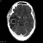
Image 6 MRI (T1 C+) ( update )

Image 7 MRI (T1 C+) ( update )

Image 8 MRI (T1 C+ fat sat) ( update )


 Unable to process the form. Check for errors and try again.