
Osteochondritis dissecans (OCD) is the end result of the aseptic separation of an osteochondral fragment with the gradual fragmentation of the articular surface and results in an osteochondral defect. It is often associated with intra-articular loose bodies.
On this page:
Epidemiology
Onset is between childhood and young adulthood, with the majority of patients being between 10 and 40 years of age. There is a male to female ratio of ~2:1 3.
Risk factors
repetitive throwing / valgus stress and gymnastics / weight-bearing on upper extremity: valgus stress / compressive force on the vulnerable chondroepiphysis of the radiocapitellar joint in skeletally immature patients is supported as the etiology for osteochondritis dissecans of the capitellum 8
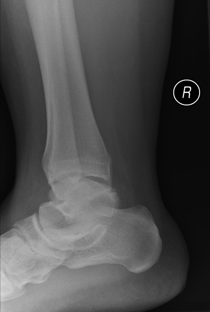
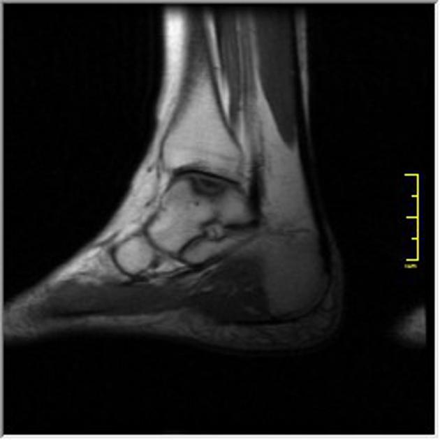
ankle sprain/instability: in the talus, 96% of lateral lesions and 62% of medial lesions are associated with direct trauma 9
competitive athletics 10
family history: epiphyseal dysplasia has been postulated as a subset of osteochondritis dissecans 11
Clinical presentation
Symptoms are variable and range from asymptomatic to significant pain and locking (suggesting loose body formation). Joint effusions and synovitis are often present.
Pathology
Etiology
The exact etiology of osteochondritis dissecans is uncertain and controversial, with the majority of cases thought to be the result of trauma 4. In up to 40% of cases, patients give a history of trauma as the inciting event 3. Other postulated causes include 4:
microtrauma
familial dysplasia
Location
Many joints can be affected, but typical locations include:
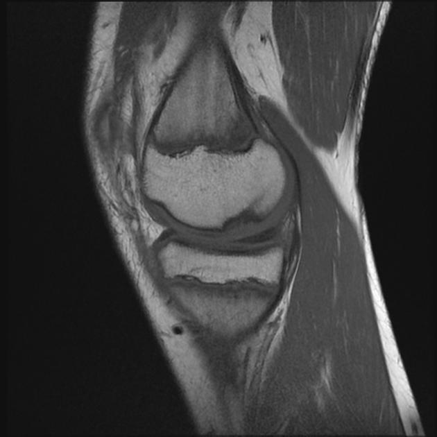
femoral condyles (most common site; ~95% of all cases): osteochondritis dissecans of the knee
glenohumeral joint: rare; humeral head more commonly involved than the glenoid 7,19
Staging
See osteochondral injury staging and osteochondritis dissecans surgical staging.
Radiographic features
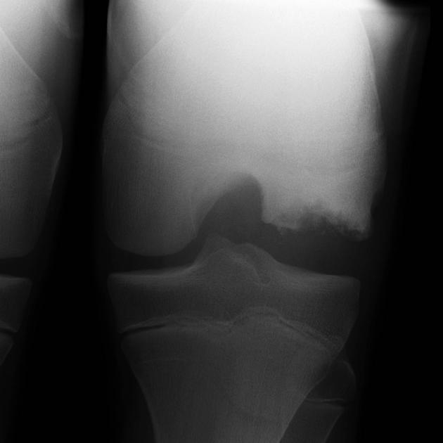
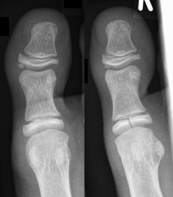
Plain radiograph
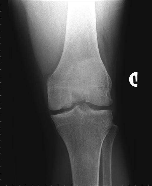
Plain radiographs should be the first step in the evaluation of knee pain, however, unless advanced changes are present and/or a meticulous technique is employed, early findings of osteochondritis dissecans may be occult. The intercondylar "notch" view is very helpful.
Early findings include subtle flattening or indistinct radiolucency above the cortical surface. As the process progresses, more pronounced contour abnormalities, fragmentation, and density changes (both lucency and sclerosis) become evident. If an osteochondral fragment becomes unstable and displaced, a donor site and intra-articular fragment may be seen.
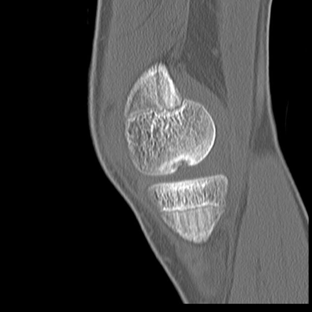
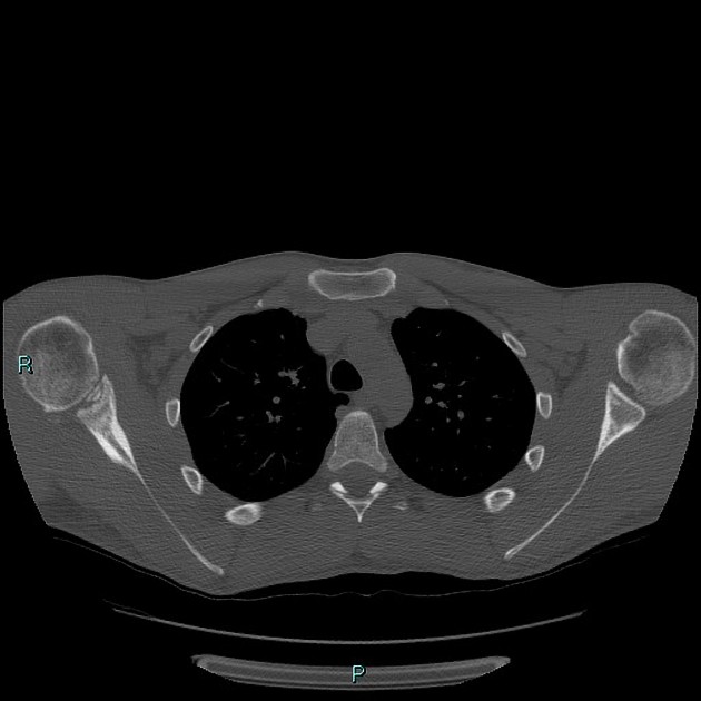
CT
CT has the advantage of sectional imaging through the joint and multiplanar reformats. Findings are similar to those seen on plain radiographs.
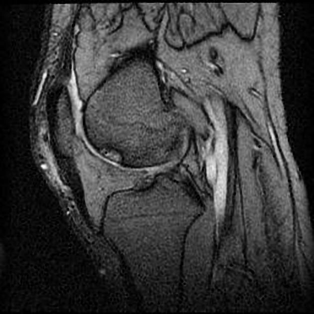
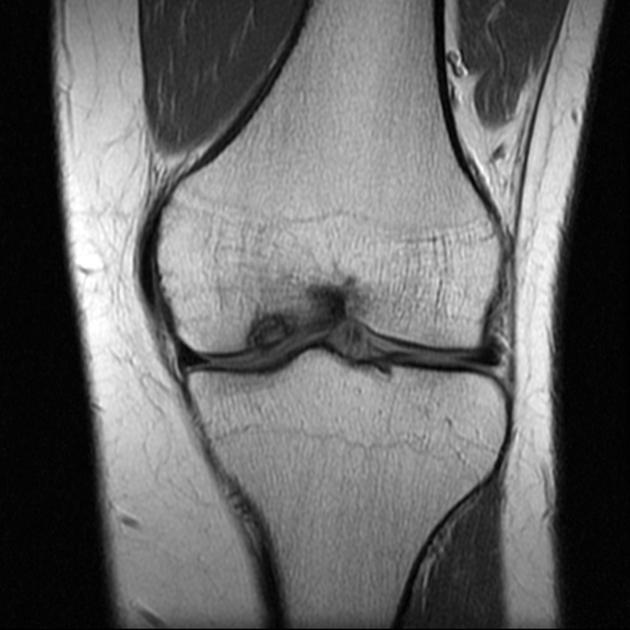
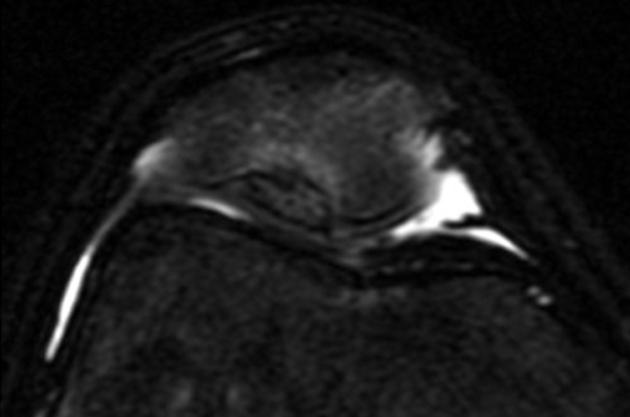
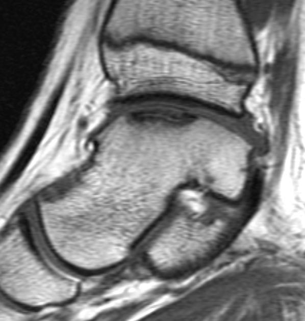
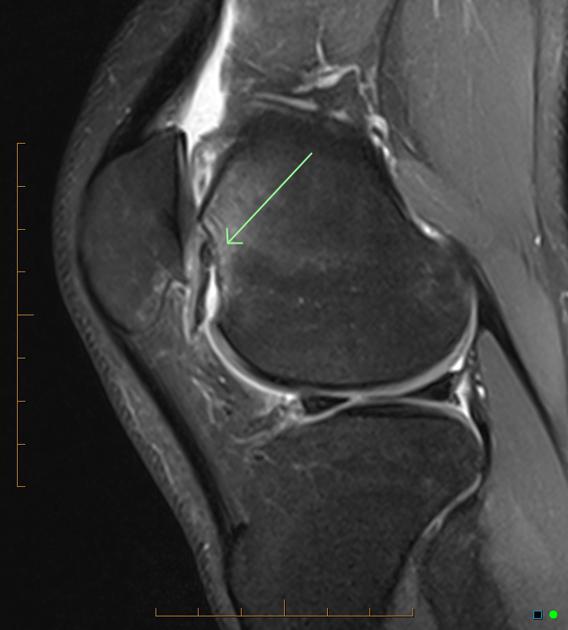
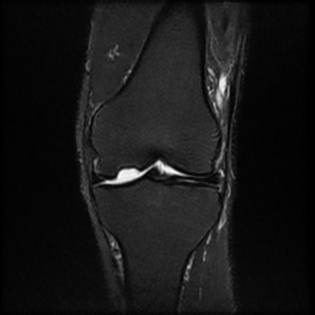
MRI
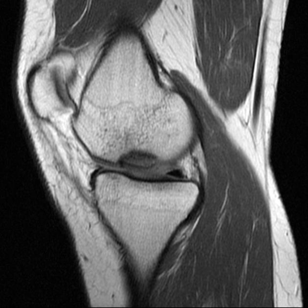
MRI is the modality of choice, with high sensitivity (92%) and specificity (90%) 4 in the detection of separation of the osteochondral fragment. This is essential in determining management. However, the actual defect may or may not be present on MR imaging depending on the stage of the process.
T1: variable signal overall with intermediate to low signal adjacent to fragment and variable fragment signal
-
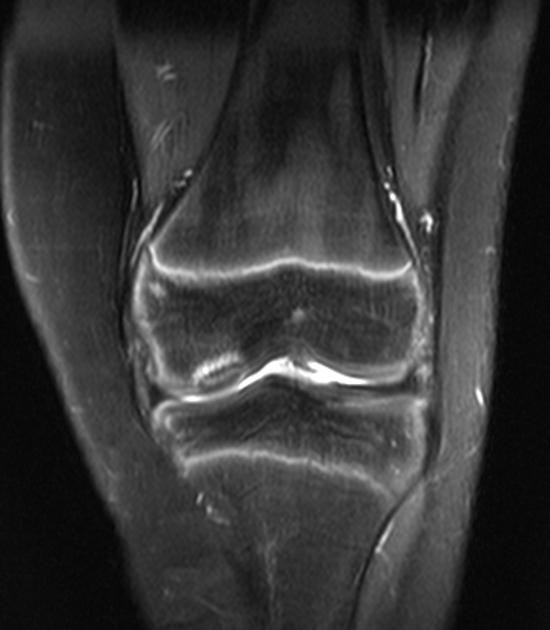
T2
the high signal line demarcating fragment from bone usually indicates an unstable lesion however false positives can result from edema 6
low signal loose bodies, outlined by high signal fluid
donor defect filled with high signal fluid
high signal subchondral cysts
T1 C+ (Gd): enhancement indicates the viability of the lesion
The four classic signs of instability described on MRI include 14:
high signal intensity rim at the interface between the fragment and the adjacent bone on T2-weighted sequences
fluid-filled cysts beneath the lesion
high signal intensity line extending through the articular cartilage overlying the lesion
focal osteochondral defect filled with joint fluid, indicating complete detachment of the fragment
Treatment and prognosis
Spontaneous healing is usual unless there is an unstable fragment, and treatment revolves around rest and immobilization for up to a year 5.
When the fragment is unstable or displaced, without treatment patients are susceptible to premature secondary osteoarthritis. Numerous surgical approaches have been tried, including drilling, bone grafting, replacement of bone fragment and pinning 5.
When surgery is performed, the results in most cases are only "fair". Around 50% (range 35-70%) of patients achieve a "good to excellent" clinical outcome 3 but even in these cases, the majority develop osteoarthritis.
Complications
persistent pain with activity: occurs in ~66% following surgical management of the knee and in 40% following surgical management of the elbow 12,13
articular incongruity 13
early degenerative joint disease 13
History and etymology
Osteochondritis dissecans was first described by the German orthopedic surgeon Franz Konig (1832-1910) 17 in 1887, and he coined the term which appeared for the first time in this article 18.


 Unable to process the form. Check for errors and try again.