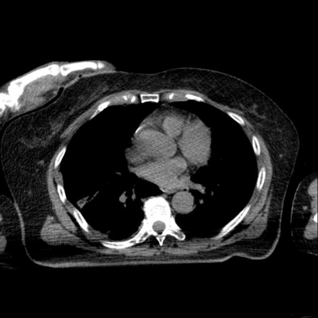
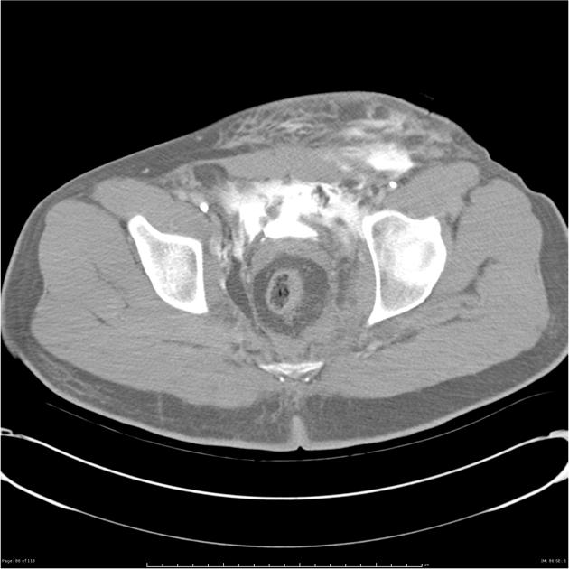
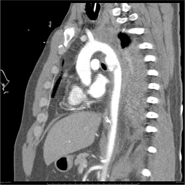
CT polytrauma/multitrauma, also called trauma CT, whole body CT (WBCT) or panscan, is an increasingly used investigation in patients with multiple injuries sustained after significant trauma.
The majority of the evidence regarding whole-body CT is, understandably, retrospective. There is some evidence from meta-analyzes that trauma patients who undergo WBCT have better survival than patients who undergo selective imaging 3,10 but this is yet to be definitely proven in randomized controlled trials 5,8,9.
Patients who undergo immediate WBCT at presentation have a similar radiation dose at discharge to patients who undergo a selective imaging strategy 9,10. As well as traumatic injuries, trauma CT uncovers incidental findings which have varying levels of significance 6,7.
On this page:
Indications
Indications will vary from institution-to-institution but indications by mechanism include:
high speed motor vehicle collision
non-trivial motorcycle collision
death at the scene
fall from height >2 meters
other concerning mechanism of injury
abnormal FAST, or trauma chest or pelvis x-ray
abnormal vital signs
Purpose
Clinical assessment and mechanism of injury may underestimate injury severity by 30% 8. The purpose of the scan is first and foremost, the rapid evaluation of life threatening injuries and secondly the accurate diagnosis of known and unknown injuries.
Technique
Standard whole-body CT
The actual procedure will vary depending on institutional protocol/guidelines, but a typical protocol will consist of:
-
contrast-enhanced chest (arterial phase)
scan extent to mid abdomen
Additions to the whole-body CT protocol
Additionally, depending on the injuries present and especially if the images are reviewed with the patient on the CT table, the following phases may be useful:
-
delayed phase of the abdomen/pelvis
useful to assess for contrast pooling/contrast extravasation indicative of active bleeding
-
angiogram from the aortic arch to vertex
used to asses penetrating neck injuries or risk factors for blunt cerebrovascular injuries
-
angiogram of the head
used to asses abnormalities of the circle of Willis
-
renal excretory phase of the abdomen/pelvis
useful in patients with traumatic renal injuries (to assess for urinoma and upgrade AAST grading)
-
to assess for bladder injury
Variations to the whole-body CT protocol
-
addition of a non-contrast chest and abdomen
used in the context of suspected bleeds
additional non-contrast CT of the upper abdomen
CT angiogram of the abdomen/pelvis and lower limbs in the setting of suspected major hemorrhage and/or pelvic/lower limb fractures
triphasic injection single pass CT of the chest, abdomen and pelvis 4


 Unable to process the form. Check for errors and try again.