Foot (weight-bearing lateral view)
Citation, DOI, disclosures and article data
At the time the article was created Jeremy Jones had no recorded disclosures.
View Jeremy Jones's current disclosuresAt the time the article was last revised Henry Knipe had no recorded disclosures.
View Henry Knipe's current disclosures- Foot weightbearing horizontal beam lateral
- Foot (weightbearing lateral view)
The weight-bearing lateral foot radiograph is important in the assessment of foot alignment and the diagnosis of abnormalities that cause malalignment and foot pain. Nonweightbearing views (e.g. lateral foot) would be inadequate for the assessment of alignment as the bones of the feet are not in their functional state.
On this page:
Indications
This view is key to the assessment of foot alignment and the diagnosis of abnormalities causing malalignment and foot pain, i.e. Lisfranc injury. This view may also be crucial in determining the integrity of any functional joint prior to a partial foot amputation or corrective surgery for pes planus.
Patient position
- patient stands on a stable, raised radiolucent platform (e.g. using wooden blocks) that matches the x-ray tube's lowest height
- lateral aspect of foot is kept in contact with the detector
- patient applies weight on affected foot
- unaffected foot is lifted; if possible, the contralateral toes can be positioned posterior to the affected foot's calcaneus as counterbalance
Technical factors
- mediolateral projection
-
centering point
- base of metatarsals or midfoot
-
collimation
- anteriorly to skin margin of the distal phalanges
- posteriorly to skin margin of the calcaneus
- superior to the talocrural joint
-
orientation
- landscape
-
detector size
- 18 cm x 24 cm
-
exposure
- 55-60 kVp
- 3-4 mAs
-
SID
- 100 cm
-
grid
- no
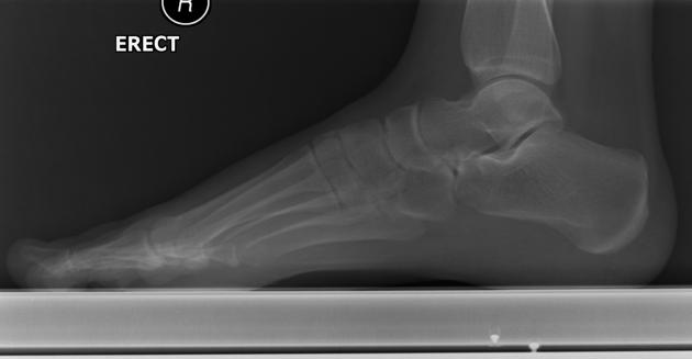
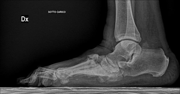
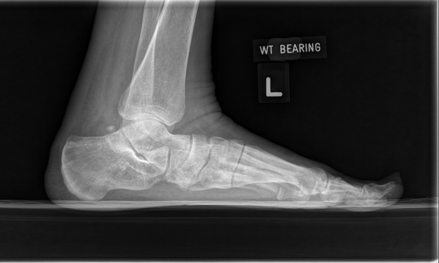
Image technical evaluation
- 5th metatarsal tuberosity is seen in profile
- the superior aspect of the talar domes are superimposed
- tibiotalar joint is open
Useful measurements
- calcaneal pitch
- medial cuneiform-fifth metatarsal height
- lateral talocalcaneal angle
- lateral talo-first metatarsal angle
- lateral calaneo-first metatarsal angle
- first metatarsal declination angle
Practical points
With patient safety being first priority, ensure some support is provided for patients to hold onto as their affected injured foot may not be used to the applied pressure.
References
- 1. John Lampignano, Leslie E. Kendrick. Bontrager's Textbook of Radiographic Positioning and Related Anatomy. (2017) ISBN: 9780323399661 - Google Books
Related articles: Imaging in practice
- imaging in practice
-
general radiography (adult)
- portable radiography
- chest radiography
- abdominal radiography
-
upper limb radiography
-
shoulder girdle radiography
- scapula series
-
shoulder series
- shoulder (AP view)
- shoulder (internal rotation view)
- shoulder (external rotation view)
- shoulder (superior-inferior axial view)
- shoulder (inferior-superior axial)
- shoulder (West Point view)
- shoulder (Velpeau view)
- shoulder (modified trauma axial view)
- shoulder (supine lateral view)
- shoulder (modified transthoracic supine lateral)
- shoulder (lateral scapula view)
- shoulder (AP glenoid view)
- shoulder (Garth view)
- shoulder (outlet view)
- shoulder (Stryker notch view)
- acromioclavicular joint series
-
clavicle series
- clavicle (AP view)
- clavicle (AP cephalic view)
- clavicle (oblique view)
- sternoclavicular joint series
- arm and forearm radiography
- wrist and hand radiography
- wrist series
- scaphoid series
- hand series
- thumb series
- fingers series
- rheumatology hands series
- bone age (radiograph)
-
shoulder girdle radiography
-
lower limb radiography
- pelvic girdle radiography
- thigh and leg radiography
- ankle and foot radiography
- skull radiography
-
paranasal sinus and facial bone radiography
- facial bones
- mandible
- nasal bone
- zygomatic arches
- paranasal sinuses
- temporal bones
- dental radiography
- orthopantomography
- temporomandibular joints
- temporomandibular joint (AP axial view)
- temporomandibular joint (axiolateral oblique view)
-
spinal radiography
- cervical spine series
-
thoracic spine series
- thoracic spine (AP view)
- thoracic spine (lateral view)
- thoracic spine (oblique view)
- lumbar spine series
- sacrococcygeal radiography
- scoliosis radiography
-
pediatric radiography
- radiographic positioning terminology
- systematic radiographic technical evaluation (mnemonic)
- pediatric immobilization
- foreign body ingestion series (pediatric)
- foreign body inhalation series (pediatric)
- shunt series
- chest radiograph (pediatric)
- abdomen radiograph (pediatric)
- upper limb radiography (pediatric)
-
lower limb radiography (pediatric)
- pelvis radiograph (pediatric)
- femur series (pediatric)
- pediatric femur (AP view)
- pediatric femur (lateral view)
- leg length view
- knee series (pediatric)
- pediatric knee (AP view)
- pediatric knee (lateral view)
- tibia fibula series (pediatric)
- pediatric tibia fibula (AP view)
- pediatric tibia fibula (lateral view)
- pediatric tibia fibula (oblique view)
- ankle series (pediatric)
- foot series (pediatric)
- skull radiography (pediatric)
- spine radiography (pediatric)
-
skeletal survey
-
skeletal survey (non-accidental injury)
- torso
- pediatric chest (AP erect view)
- pediatric chest (oblique ribs view)
- pediatric abdomen (AP supine view)
- pediatric spine (whole lateral view)
- upper limb (both sides)
- lower limb (both sides)
- pediatric femur (AP view)
- pediatric knee (lateral view)
- pediatric tibia fibula (AP view)
- pediatric ankle (lateral view)
- pediatric foot (DP view)
- torso
-
skeletal survey (non-accidental injury)
-
CT
- iodinated contrast media
- CT IV contrast media administration
-
CT protocol
- composite
- whole-body CT (protocol)
- CT Chest abdomen-pelvis (protocol)
- CT NCAP (neck, chest, abdomen and pelvis)
- head & neck
- chest
- abdomen and pelvis
- CT abdomen-pelvis (protocol)
- CT abdominal aorta
- CT adrenals (protocol)
- CT cholangiography (protocol)
- CT colonography (protocol)
- CT enteroclysis (protocol)
- CT enterography (protocol)
- CT gastrography (protocol)
- CT kidneys, ureters and bladder (protocol)
- CT urography (protocol)
- CT Renal mass (protocol)
- CT angiography of the splanchnic vessels (protocol)
- CT renal split bolus
- CT pancreas (protocol)
- liver
- composite
- barium studies
-
MRI
- cine imaging
-
brain
- screen protocol
- stereotaxis protocol
- tumor protocol
- stroke protocol
- infection protocol
- trauma protocol
- demyelination protocol
- epilepsy protocol
- neurodegenerative protocol
- trigeminal neuralgia protocol
- posterior fossa protocol
- temporal bone/IAM/CPA protocol
- pineal and tectal plate protocol
- pituitary gland protocol
- CSF flow
- angiographic protocols
- MRA
- circle of Willis (COW)
- carotid-vertebral system
- MRV
- MRA
- head and neck
- orbits protocol
- sinonasal tract protocol
- salivary glands protocol
- oropharynx and oral cavity protocol
- suprahyoid neck protocol
- infrahyoid neck protocol
- chest
- mediastinum
- cardiac
- abdomen and pelvis
- abdomen
- liver and biliary tree
- liver protocol
- MRCP: cholangiopancreatography
- pancreas protocol
- elastography
- anus and rectum
- uterus and ovaries
- urological
- prostate cancer protocol
- bladder cancer protocol
- kidneys protocol
- musculoskeletal
-
ultrasound
- ultrasound signs
-
obstetric ultrasound
- other
- placenta
-
second trimester
- fetal biometry
- fetal morphology assessment
-
soft markers
- nuchal fold thickness
- ventriculomegaly
- absent nasal bone
- echogenic intracardiac focus
- choroid plexus cysts
- echogenic bowel
- aberrant right subclavian artery
- amnioreduction
- echogenic fetal bowel
- umbilical artery Doppler assessment
- chorionic villus sampling (CVS) and amniocentesis
- nuchal translucency
- failed early pregnancy
- subchorionic hematoma
- multiple gestations
- ectopic pregnancy
-
first trimester and early pregnancy
- gestational sac
- yolk sac
- embryo/fetus
- amnion
- chorion
- Beta-hCG levels
- gynecologic ultrasound
- vascular ultrasound
- carotids
- extremities
- mesenteric vessels
- other
- breast ultrasound
-
musculoskeletal ultrasound
- technique/artifacts
- ultrasound of arthropathies
- skin/soft tissue ultrasound
- lipoma
- parasitic infection
- other
- pediatric musculoskeletal ultrasound
- ankle/foot ultrasound
-
knee ultrasound
- Baker cyst (popliteal cyst)
- infrapatellar bursitis
- hip ultrasound
- hand ultrasound
- wrist ultrasound
- elbow ultrasound
- shoulder ultrasound
- liver ultrasound
- hyperechoic liver lesion
- sonographic halo sign
- ultrasound appearances of liver metastases
- periportal hyperechogenicity
- periportal hypoechogenicity
- generalized increase in hepatic echogenicity
- generalized reduced hepatic echogenicity
- coarsened hepatic echotexture
- starry sky appearance (ultrasound)
- normal hepatic vein Doppler
- hepatic arterial resistive index
- gallbladder ultrasound
- pancreatic ultrasound
- gastrointestinal ultrasound
- renal ultrasound
- bladder ultrasound
-
testicular and scrotal ultrasound
- other
- epididymis
- paratesticular lesions
- bilateral testicular lesion
-
unilateral testicular lesion
- testicular torsion
- orchitis
- testicular rupture
-
germ cell tumor of the testis
- testicular seminoma
-
non seminomatous germ cell tumors
- mixed germ cell tumor
- yolk sac tumor (endodermal sinus tumor)
- embryonal cell carcinoma
- choriocarcinoma
- testicular teratoma
- testicular epidermoid (teratoma with ectodermal elements only)
- burned out testis tumor
- sex cord / stromal tumors of the testis
- prostate ultrasound
- neck and thyroid ultrasound
- echocardiography
- speckle tracking echocardiography
- fetal echocardiography
- contrast-enhanced echocardiography
- epicardial echocardiography
- three dimensional (3D) echocardiography
- transesophageal echocardiography (TEE)
- transthoracic echocardiography (TTE)
- left ventricular systolic and diastolic function
- structure and morphology
- systolic function
- diastolic function
- right ventricular assessment
- right and left atria
- valvular structure and function
- mitral valve
- aortic valve
- pulmonic valve
- tricuspid valve
- hemodynamics
- pericardium
- cardiomyopathies
- congenital heart disease
- great vessels
- pediatric ultrasound
- ultrasound interventions
- ultrasound-guided biopsy
- ultrasound-guided percutaneous drainage
- ultrasound-guided musculoskeletal interventions
- joint injection
- nerve blocks
- ultrasound-guided intravenous cannulation
- contrast-enhanced ultrasound
- physics and imaging modes
- grey-scale (B-mode)
- motion mode (M-mode)
- color flow Doppler (CFD)
-
spectral Doppler
- pulsed wave Doppler (PWD)
- continuous wave Doppler (CWD)
- superb microvascular imaging (SMI)
- tissue Doppler imaging (TDI)
- nuclear medicine
-
radiation therapy
- external beam radiation therapy (EBRT)
- sealed source radiation therapy (brachytherapy)
- unsealed source radiation therapy
-
interventional
- procedure overview
- neck
- thyroid gland
- breast
- chest
- hepatobiliary
- splenic interventions
- gastrointestinal
- urogenital
- bladder
- kidney
- prostate
- musculoskeletal
-
arthrogram
- MR arthrogram
- CT arthrogram
- anesthetic arthrogram
- bone biopsy (CT-guided)
-
arthrogram
- upper limb
- lower limb
-
hip
- hip joint injection (technique)
- greater trochanteric bursa injection
- gluteus minimus/medius tendon calcific tendinopathy barbotage
- iliopsoas tendon bursa injection
- lateral cutaneous femoral nerve of the thigh injection
- piriformis injection
- common hamstrings origin injections
-
knee
- common peroneal (fibular) nerve injection
- knee joint injection
- patella tendon microtenotomy
- quadriceps tendon microtenotomy
- tibial nerve injection
-
ankle
- achilles hydrodilation & microtenotomy
- ankle joint injection
- extensor tendon sheath injection
- flexor tendon sheath injection
- os trigonum injection
- peroneal tendon sheath injection
- subtalar joint injection
- tibial nerve injection
-
foot
- calcaneocuboid joint injection
- metatarsophalangeal joint (MTPJ) injection
- naviculocuneiform joint injection
- plantar fascia microtenotomy
- subtalar joint injection
- talonavicular joint injection
- tarsometatarsal joint (TMTJ) injection
-
hip
- spine
- breast imaging
-
mammography
- breast screening
- breast imaging and the technologist
- forbidden (check) areas in mammography
-
mammography views
- craniocaudal view
- mediolateral oblique view
- additional (supplementary) views
- true lateral view
- lateromedial oblique view
- late mediolateral view
- step oblique views
- spot view
- double spot compression view
- magnification view
- exaggerated craniocaudal (axillary) view
- cleavage view
- tangential views
- caudocranial view
- bullseye CC view
- rolled CC view
- elevated craniocaudal projection
- caudal cranial projection
- 20° oblique projection
- inferomedial superolateral oblique projection
- Eklund technique
- normal breast imaging examples
-
mammography
- digital breast tomosynthesis
- breast ultrasound
- breast ductography
- breast MRI
- breast morphology
- breast intervention