
Meniscal tears are the failure of the fibrocartilaginous menisci of the knee. There are several types and can occur in an acute or chronic setting. Meniscal tears are best evaluated with MRI.
On this page:
Pathology
Acute meniscal tears occur after the rotatory trauma of the knee, whereas chronic degenerative meniscal tears often occur in the elderly after minimal rotatory trauma or stress on the knee.
In older adults, attritional changes in the meniscus lead to fragmentation of the meniscus and a variety of tears (usually occur at the posterior horn of the medial meniscus) 8.
Types
There are different types of meniscal tears, describing the morphology of the injury. Identifying and accurately describing the type of meniscal tear can help the surgeon in patient education and planning of the surgical procedure. Meniscal tear types include 1,5,6:
-
basic tears
-
longitudinally oriented tears
-
horizontal tear (cleavage tear)
parallel to the tibial plateau involving one of the articular surfaces or free edge
divides the meniscus into superior and inferior parts
-
perpendicular to the tibial plateau and parallel to the long axis of the meniscus
divides the meniscus into medial and lateral parts
Wrisberg rip - a specific lateral meniscus subtype
ramp lesion - a specific medial meniscus subtype
-
radial tear: perpendicular to both the tibial plateau and the long axis of the meniscus
root tear: typically radial-type tear located at the meniscal root
-
complex tear: a combination of all or some horizontal, vertical, and radial-type tears
-
displaced tear: tear involving a component that is displaced, either still attached to the parent meniscus or detached:
flap tear: displaced horizontal or vertical tears
bucket-handle tear: displaced vertical tear
parrot beak tear: oblique radial tear
Radiographic features
Plain radiograph
On plain radiographs, meniscal tears are not visible. In rare cases secondary signs can be seen, such as a soft tissue swelling next to the meniscus when a meniscal cyst is present 4. Only when associated with more complex injuries plain film may suggest a meniscal tear, e.g. arcuate sign, reverse Segond fracture, tibial plateau fracture.
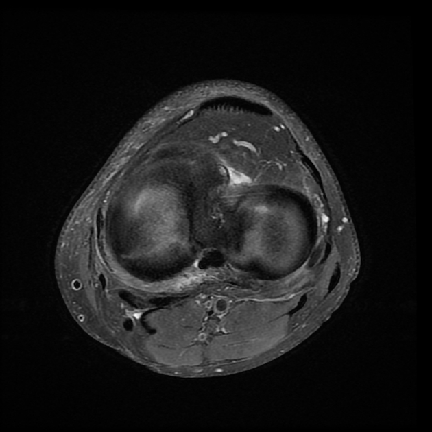
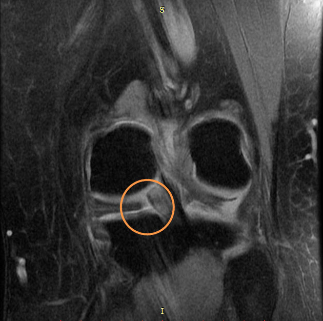
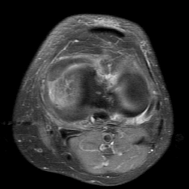
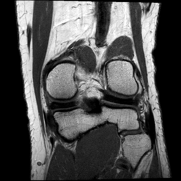
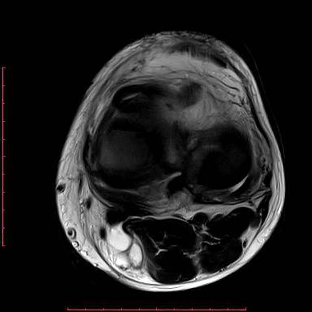
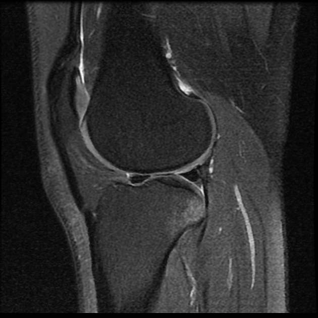
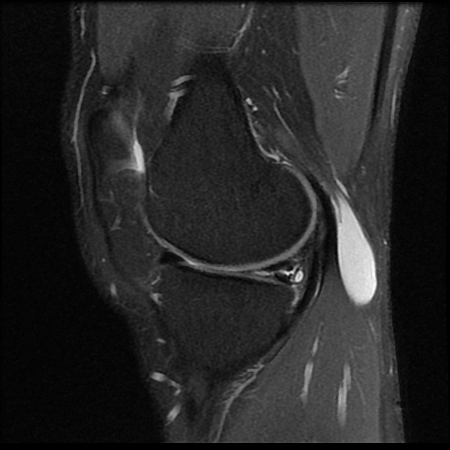
MRI
With a sensitivity of ~95% and a specificity of 81% for medial meniscal tears and sensitivity of ~85% and a specificity of 93% for lateral meniscal tears 2,5, MRI is the modality of choice when a meniscal tear is suspected, with sagittal images being the most sensitive 5.
There are three basic MRI characteristics/criteria of meniscal tears 5:
high intrameniscal signal extending to at least one articular surface, which should be seen in at least two slices: two slice touch rule (do not have to be contiguous, e.g. sagittal and coronal slices)
distortion of the normal meniscal morphology if no prior surgery
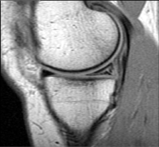
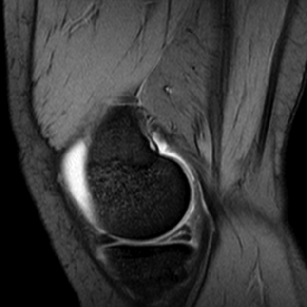
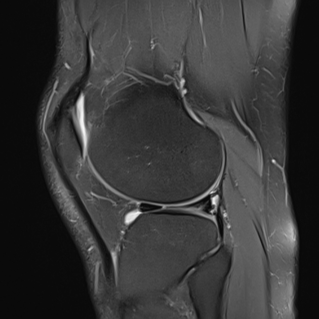
Each type of meniscal tear has its own characteristics on MRI, but in most cases, the following can be seen 3:
T1: a hyperintense line in the meniscus can be seen, but it is difficult to differentiate between degeneration and meniscal tear on this sequence; in the case of a bucket-handle tear an empty groove can sometimes be seen
-
T2: a hyperintense line in the meniscus, which indicates synovial fluid in the meniscus
-
the high T2 signal in mid-substance of the meniscus without extension to the surface is not necessarily a tear and can be:
in adults: secondary to degeneration
in children: high vascularity of meniscus
-
See MRI grading system for meniscal signal intensity.
Associated features that are suggestive of a meniscal tear include 5:
There are several signs associated with meniscal tears:
truncated triangle sign
flipped meniscus sign
quadruple sign
Treatment and prognosis
Surgical arthroscopy is done in most cases. Meniscopexy or complete or partial meniscectomy can be performed, depending on the degree and type of meniscal tear.
Practical points
There are numerous pitfalls to be aware of ref:
oblique ligament: with intercondylar bucket handle component
previous surgery
fluid in normal central knee recesses
fluid in popliteal hiatus
chondrocalcinosis: can increase signal intensity on MRI
meniscal high signal can be normal in adolescents and young adults ref
Differential diagnosis
The differential can be variable, depending on the type of tear but in general, consider:
meniscal degeneration (can be associated with a tear)
meniscal flounce (rare)
ring meniscus (rare)
meniscal ossicle (rare)


 Unable to process the form. Check for errors and try again.