
Ischemic penumbra
Citation, DOI, disclosures and article data
At the time the article was created Frank Gaillard had no recorded disclosures.
View Frank Gaillard's current disclosuresAt the time the article was last revised Arlene Campos had no financial relationships to ineligible companies to disclose.
View Arlene Campos's current disclosures- Ischemic penumbra
- Penumbra
Ischemic penumbra denotes the part of an acute ischemic stroke that is at risk of progressing to infarction but is still salvageable if reperfused. It is usually located around an infarct core which represents the tissue which has already infarcted or is going to infarct regardless of reperfusion.
The primary aim of current acute stroke intervention is to prevent the penumbra from proceeding to established infarct.
Radiographic features
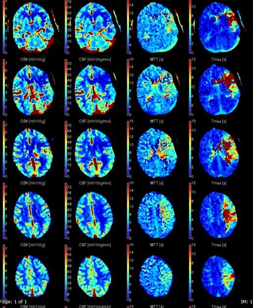
CT perfusion
On CT perfusion the ischemic penumbra has been variably defined using a combination of parameters. Generally, it should be conceptually thought of as "the area of the brain with reduced perfusion" minus the "infarct core". In practice, which parameters are used to define each component has varied. A fairly common definition for penumbra is the area of brain with 1,2:
prolonged (increased) T-max, typically >6 seconds (or other measures of delayed arrival of contrast such as mean transit time (MTT) or time to peak (TTP)), and...
normal or increased cerebral blood volume (CBV) due to autoregulation
This region will have only a moderate decreased cerebral blood flow (CBF). This is in contrast to the infarct core which will have a marked decrease in cerebral blood flow and also a decrease in cerebral blood volume.
MRI
On MRI, the ischemic penumbra is also determined by the area of the brain with reduced perfusion 1:
penumbra: shows perfusion changes same as that with CT
infarcted core: shows restricted diffusion (established infarct) apart from decreased cerebral blood volume and cerebral blood flow
Quiz questions

References
- 1. Srinivasan A, Goyal M, Al Azri F et-al. State-of-the-art imaging of acute stroke. Radiographics. 2006;26 Suppl 1 (suppl 1): S75-95. Radiographics (full text) - doi:10.1148/rg.26si065501 - Pubmed citation
- 2. de Lucas E, Sánchez E, Gutiérrez A et al. CT Protocol for Acute Stroke: Tips and Tricks for General Radiologists. Radiographics. 2008;28(6):1673-87. doi:10.1148/rg.286085502 - Pubmed
Incoming Links
- Stroke protocol (CT)
- MR perfusion weighted imaging
- Acute basilar artery occlusion
- Terms used in radiology
- Cerebral intraparenchymal hyperattenuation post thrombolysis
- CT perfusion in ischaemic stroke
- Prominent vessel sign
- CT brain perfusion (protocol)
- Ghost infarct core
- Infarct core
- Potential recuperation ratio (PRR)
- Endovascular clot retrieval (ECR)
- Ischemic stroke
- Code stroke CT (an approach)
- Central nervous system curriculum
Related articles: Stroke and intracranial haemorrhage
-
stroke and intracranial hemorrhage
- general articles
-
ischemic stroke
- general discussions
- scoring and classification systems
- Alberta stroke program early CT score (ASPECTS)
- ASCOD classification
- Canadian Neurological Scale
- Heidelberg bleeding classification
- NIH Stroke Scale
- Mathew stroke scale
- modified Rankin scale
- Orgogozo Stroke Scale
- Scandinavian Stroke Scale
- thrombolysis in cerebral infarction (TICI) scale
- TOAST classification
- collateral vessel scores
- signs
- by region
- hemispheric infarcts
- frontal lobe infarct
- parietal lobe infarct
- temporal lobe infarct
- occipital lobe infarct
- alexia without agraphia syndrome: PCA
- cortical blindness syndrome (Anton syndrome): top of basilar or bilateral PCA
- Balint syndrome: bilateral PCA
- lacunar infarct
-
thalamic infarct
- artery of Percheron infarct
- Déjerine-Roussy syndrome (thalamic pain syndrome): thalamoperforators of PCA
- top of the basilar syndrome
- striatocapsular infarct
- choroid plexus infarct
- cerebellar infarct
-
brainstem infarct
- midbrain infarct
- Benedikt syndrome: PCA
- Claude syndrome: PCA
- Nothnagel syndrome: PCA
- Weber syndrome: PCA
- Wernekink commissure syndrome
- pontine infarct
- Brissaud-Sicard syndrome
- facial colliculus syndrome
- Gasperini syndrome: basilar artery or AICA
- inferior medial pontine syndrome (Foville syndrome): basilar artery
- lateral pontine syndrome (Marie-Foix syndrome): basilar artery or AICA
- locked-in syndrome: basilar artery
- Millard-Gubler syndrome: basilar artery
- Raymond syndrome: basilar artery
- medullary infarct
- Babinski-Nageotte syndrome
- Cestan-Chenais syndrome
- hemimedullary syndrome (Reinhold syndrome)
- lateral medullary stroke syndrome (Wallenberg syndrome)
- medial medullary syndrome (Déjerine syndrome)
- Opalski syndrome
- midbrain infarct
- acute spinal cord ischemia syndrome
- hemispheric infarcts
- by vascular territory
- by vessel size
- treatment options
- complications
-
intracranial hemorrhage
-
intra-axial hemorrhage
- signs and formulas
- ABC/2 (volume estimation)
- black hole sign
- blend sign
- cashew nut sign
- CTA spot sign
- island sign
- satellite sign
- swirl sign
- zebra sign
- by type
- by location
- signs and formulas
- extra-axial hemorrhage
- extradural hemorrhage (EDH)
- intralaminar dural hemorrhage
- subdural hemorrhage (SDH)
-
subarachnoid hemorrhage (SAH)
- types
- complications
- grading systems
- subpial hemorrhage
-
intra-axial hemorrhage
Related articles: Terms used in radiology
- general
- ancillary
- Cinderella
- diagnosis of exclusion
- dilation vs dilatation
- epiphenomenon
- florid
- forme fruste
- gold standard
- heterogeneous vs heterogenous
- Hickam's dictum
- iatrogenic disease
- idiopathic
- in extremis
- natural history
- non-specific
- Occam's razor
- prodrome
- Saint's triad
- self-limiting
- sequela
- sine qua non
- status post
- subclinical disease
- syndrome
- radiology-specific
- pathology
- agenesis
- anlage
- aplasia
- apoptosis
- atresia
- atrophy
- cyst
- dehiscence
- diathesis
- diverticulum
- dyscrasia
- dysplasia
- exophytic
- fistula
- fluid collection
- granulation tissue
- hernia
- hyperplasia
- hypertrophy
- hypoplasia
- lamellated
- laminated
- malignancy
- metaplasia
- necrosis
- neoplasm
- phlegmon
- septum
- synechia
- trabecula
- CNS
- chest
- epidemiology
- gastrointestinal
- genetics
- musculoskeletal
- oncology


 Unable to process the form. Check for errors and try again.