
Ossifying fibromas are benign bone lesions that should be differentiated from non-ossifying fibromas and fibrous dysplasia. Osteofibrous dysplasia is considered as a separate pathological entity in view of its different presentation and treatment, although histopathologically similar to ossifying fibroma.
On this page:
Epidemiology
These lesions are most frequently encountered in young children (often <10 years).
Associations
Pathology
Location
-
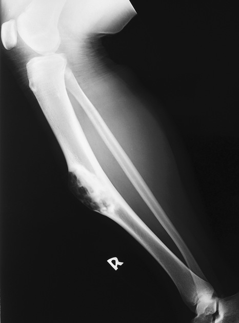
lower extremity
tibia: most frequent site 5 (90% of the time); there is a predilection for the anterior tibial cortex
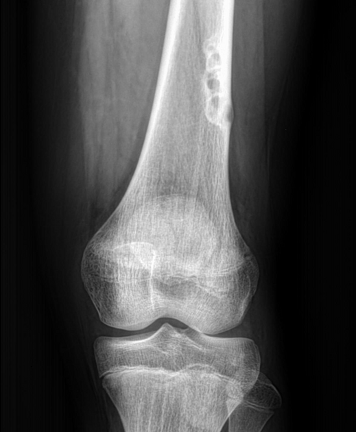
femur: occurs in a diaphysial location
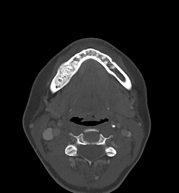
mandible and maxilla: these are examples of cementum-poor cemento-ossifying fibromas 2 (see WHO classification scheme for odontogenic tumors)
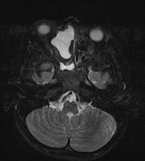
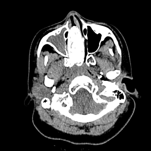
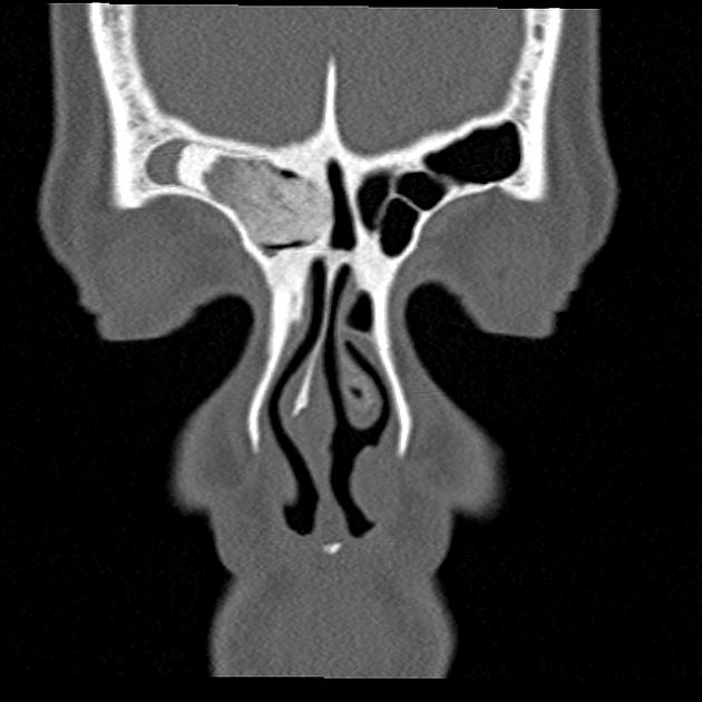
sinonasal: expansile lesions with peripheral ossification and central lucency
Microscopic appearance
They comprise haphazardly distributed lamellated bony spicules on a background of fibrous stroma. Despite being benign, they can be locally aggressive.
Immunophenotype
Immunohistochemical staining of lesions shows positive keratin cells in the majority of the cases.
Radiographic features
Plain radiograph / CT
well-circumscribed lesion
evidence of intracortical osteolysis with a characteristic sclerotic band (osteoblastic rimming)
moderate cortical expansion
homogeneous lesion matrix
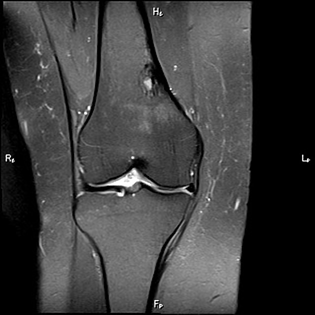
MRI
Reported signal characteristics include:
T1: low signal
T2: iso-high signal; fluid-fluid levels may be present 8
T1 C+ (Gd): typically shows enhancement
Treatment and prognosis
Ossifying fibromas tend to regress over time. For locally aggressive lesions, surgical resection is often curative although recurrence has been reported.
Complications
Differential diagnosis
Imaging differential considerations include:
fibrous dysplasia: has no osteoblastic rimming
adamantinoma: may share a common origin with ossifying fibromas


 Unable to process the form. Check for errors and try again.