
Clear cell chondrosarcomas are low-grade malignant chondrogenic bone neoplasms of the epiphysis. The name derives from the presence of clear cell chondrocytes which contain abundant vacuolated cytoplasm due to the presence of glycogen.
On this page:
Epidemiology
Clear cell chondrosarcomas make up around 2% of all chondrosarcomas 1,2 and are usually seen in the 3rd to 5th decade even though they have been reported in an age range of 12-84 years 1. Men are almost three times as often affected as women 1.
Diagnosis
The diagnosis of clear cell chondrosarcomas is mainly based on typical pathology 1.
Diagnostic criteria
The diagnostic criteria are according to the WHO classification of soft tissue and bone tumors (5thedition) 1. The essential criteria are 1:
clear cells with abundant cytoplasm and central nuclei
woven bone and osteoclastic giant cells
An epiphyseal location of the tumor is a desirable criterion 1.
Clinical presentation
Clinically, clear cell sarcomas most commonly present with pain. Symptoms persist for an average duration of one year or up to five years, suggesting slow growth of the lesion 1.
Pathology
Clear cell chondrosarcomas are low-grade malignant cartilaginous bone neoplasms often found in the epiphyses and characterized by the presence of lobules of clear cells that contain an abundant clear or mildly eosinophilic cytoplasm with centrally placed nuclei 1-3.
Etiology
The etiology of clear cell chondrosarcomas is not known 1.
Location
The lesion is most often in an epi-metaphyseal location of long tubular bones 1-4 (in contradistinction to conventional chondrosarcoma, which is usually metaphyseal-diaphyseal). Yet, they have been described in several locations of the skeleton including the skull, spine, ribs, hands and feet 1-4.
proximal femur: 55-60% (most common)
proximal humerus: 10-15%
Macroscopic appearance
Grossly, clear cell chondrosarcomas usually contain soft gritty material with features of hyaline cartilage; the latter may be focal or absent 1.
Microscopic appearance
Histologically, clear cell chondrosarcomas are characterized by the following 1-4:
lobules of cells with abundant clear or mildly eosinophilic cytoplasm
distinct cytoplasmic membranes
large round nuclei with prominent central nucleoli
only mild atypia, rare mitoses
reactive (woven) bone formation
intermingled osteoclastic giant cells
calcified or ossified hyaline cartilage
areas of low-grade chondrosarcoma may be present
possible aneurysmal bone cyst-like changes
Immunophenotype
On immunohistochemistry, clear cells express S100 1,4-6 and show reactivity for collagen types II and X 1. They can also be positive for cytokeratins 1.
Radiographic features
Clear cell chondrosarcomas are usually well-defined osteolytic lesions often found in the epi-metaphysis of the proximal femur or humerus 1-3. A diaphyseal extension is rare 7. They might have matrix mineralization, thin sclerotic borders or cystic changes 2-4,7.
Lesions in the proximal humerus usually appear more aggressive than in other long bones 2,7.
Lesions in the axial skeleton are typically more expansive, lack mineralization and tend to show soft tissue extension more often than their counterparts in the long bones 2.
Plain radiograph
On radiographs, clear cell chondrosarcomas are predominantly radiolucent and expansile 2,3,5-7:
endosteal irregularity is not infrequent
a periosteal reaction is usually absent
soft tissue extension and cortical break are usually seen only if a pathologic fracture occurs
a calcified matrix might be present
a peripheral sclerotic rim is common (simulating a benign lesion)
variable transition zone
They are usually classified as grade 1A or 1B on the Lodwick classification of lytic bone lesions 6.
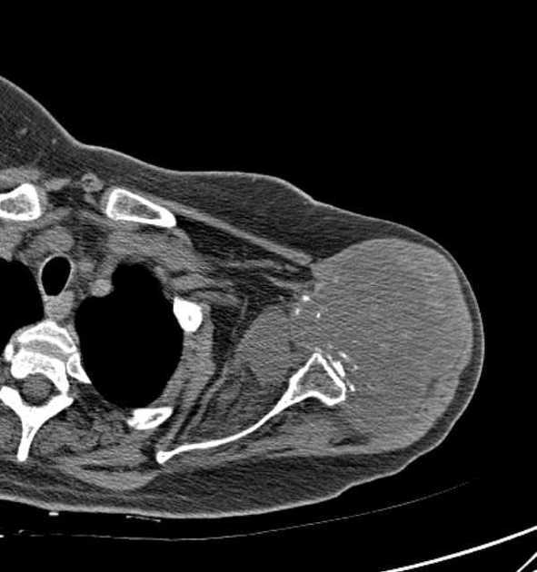
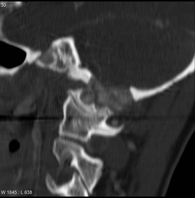
CT
Clear cell chondrosarcomas may appear as solitary lucent bone lesions on CT 2.
Matrix mineralization with a chondroid matrix as well as concerning features such as cortical involvement and soft tissue extension can be more readily depicted 2.
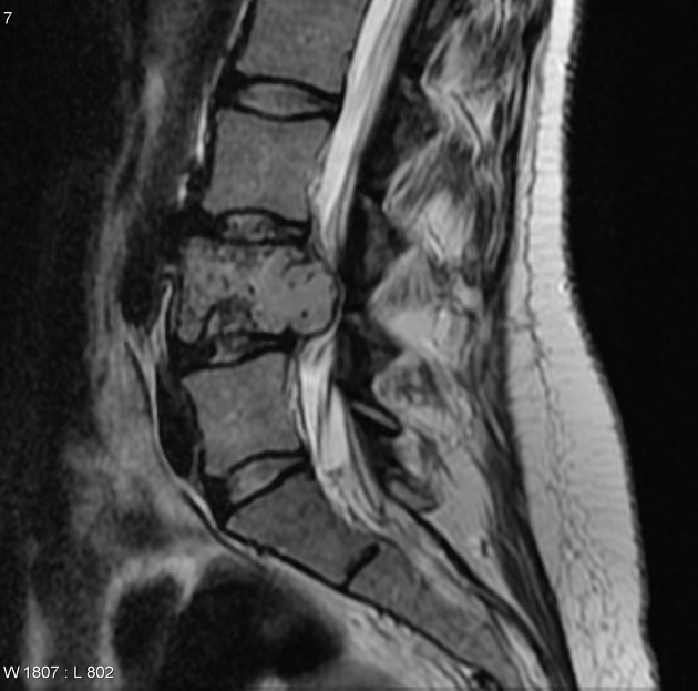
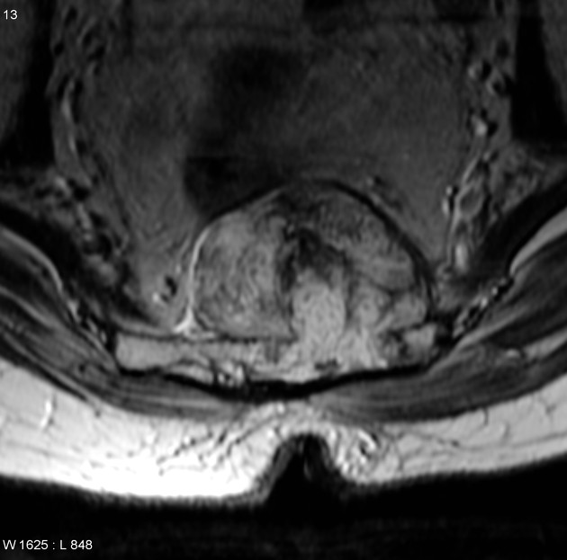
MRI
On MRI clear cell chondrosarcomas are mostly characterized as low T1 and high T2 bone lesions with signal heterogeneity due to matrix mineralization or intralesional hemorrhage 2,7.
Surrounding perilesional bone marrow edema is absent or minimal in most cases 2,7.
Typically reported signal characteristics on MR imaging include 1,2,5-7:
T1: low signal intensity
T2: moderate to high signal intensity
T1 C+ (Gd): diffuse or heterogeneous enhancement
Radiology report
The radiological report should include a description of the following 8:
location and size
tumor margins and transition zone
relation to the growth plate and articular surface
cystic and solid tumor components
fluid-fluid levels suggesting aneurysmal bone cyst-like changes
-
additional features
cortical involvement
soft tissue extension
periosteal reaction
surrounding bone marrow edema
solid mass-like enhancement
On CT and MRI the lesion can be categorized as Bone-RADS 4 unless histology has been already obtained 8.
Treatment and prognosis
Clear cell chondrosarcomas are relatively slow-growing. The treatment of choice is en bloc resection, which is usually curative with clear margins 1,2,9, whereas marginal excision and curettage are associated with high recurrence rates 1,2. Bone and lung metastases occur in 15-20% 1,9, and the overall mortality is about 15% 1,6, therefore surveillance of 10 years or longer has been advocated 9.
Complications
Complications include 2,3:
tumor recurrence
metastases to lung and bone (in exceptional cases)
History and etymology
Clear cell chondrosarcomas were first described by the American pathologist K Krishnan Unni and his research colleagues John W Beabout, David Carl Dahlin and Franklin H Sim in 1976 2-5,10.
Differential diagnosis
The differential diagnosis includes lesions with a predilection for the epiphysis (see differential for an epiphyseal lesion). Specific lesions that need to be considered include the following 1:
chondroblastoma: a particular feature of clear cell chondrosarcomas is a predilection for the epiphysis, in which case they mimic chondroblastomas; distinguishing between the two can be difficult (see chondroblastoma vs. clear cell chondrosarcoma)
lytic bone metastases (especially from clear cell renal cell carcinoma)


 Unable to process the form. Check for errors and try again.