
Adductor magnus muscle
Citation, DOI, disclosures and article data
At the time the article was created a b had no recorded disclosures.
View a b's current disclosuresAt the time the article was last revised Daniel J Bell had no recorded disclosures.
View Daniel J Bell's current disclosures- Adductor magnus muscles
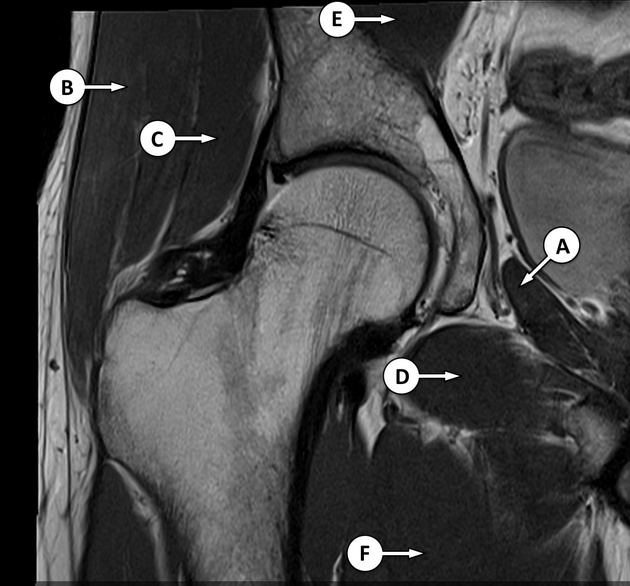
The adductor magnus muscle is the largest and deepest of the muscles in the medial compartment of the thigh. Like the adductor longus and brevis muscles, the adductor magnus is a triangular or fan-shaped muscle anchored by its apex to the pelvis and attached by its expanded base to the femur.
On this page:
Summary
-
origin
- adductor part: ischiopubic ramus
- hamstring part: ischial tuberosity
-
insertion
- adductor part: posterior surface of proximal femur, linea aspera, medial supracondylar line
- hamstring part: adductor tubercle and supracondylar line
- action: adducts and medially rotates thigh at hip joint
- arterial supply: profunda femoris
-
innervation
- adductor part: obturator nerve (L2-L4)
- hamstring part: sciatic nerve (L4-S1)
Gross anatomy
Relations
Near the base of the adductor magnus muscle is the adductor hiatus which is the terminal portion of the adductor canal 4. The contents of the adductor canal include the superficial femoral artery and vein, nerve to vastus medialis and saphenous nerve. When the superficial femoral artery and vein exit the adductor hiatus and enter the popliteal fossa they become the popliteal artery and vein respectively.
Related pathology
Quiz questions
References
- 1. Gray's Anatomy for Students: With STUDENT CONSULT Online Access, 3e. Churchill Livingstone. ISBN:0702051314. Read it at Google Books - Find it at Amazon
- 2. Clinically Oriented Anatomy. Lippincott Williams & Wilkins. ISBN:1451119453. Read it at Google Books - Find it at Amazon
- 3. Last's Anatomy. Churchill Livingstone. ISBN:0702033944. Read it at Google Books - Find it at Amazon
- 4. Leon Chaitow, Judith DeLany. Clinical Application of Neuromuscular Techniques, Volume 2. (2019) ISBN: 9780443068157
- 5. Flavia De Oliveira, Ricardo Bragança De Vasconcellos Fontes, Josemberg Da Silva Baptista, William Paganini Mayer, Silvia De Campos Boldrini, Edson Aparecido Liberti. The connective tissue of the adductor canal – a morphological study in fetal and adult specimens. (2009) Journal of Anatomy. 214 (3): 388. doi:10.1111/j.1469-7580.2009.01047.x - Pubmed
- 6. Zhou Y, Ryer EJ, Garvin RP, Irvan JL, Elmore JR. Adductor canal compression syndrome in an 18-year-old female patient leading to acute critical limb ischemia: A case report. (2017) International journal of surgery case reports. 37: 113-118. doi:10.1016/j.ijscr.2017.06.030 - Pubmed
Incoming Links
- Knee (horizontal beam lateral view)
- Duchenne muscular dystrophy
- Obturator nerve neuropathy
- Medial compartment of the thigh
- Sciatic nerve motor distribution
- Knee (lateral view)
- Adductor minimus muscle
- Inferior pubic ramus
- Adductor canal
- Vastus medialis muscle
- Sciatic neuropathy
- Adductor longus muscle
- Becker muscular dystrophy
- Descending genicular artery
- Profunda femoris artery
- Muscles of the lower limb
- Femoral artery
- Cortical desmoid
- Linea aspera
- Adductor canal syndrome
Related articles: Anatomy: Lower limb
- skeleton of the lower limb
- joints of the lower limb
-
hip joint
- ligaments
- muscles
- additional structures
- hip joint capsule
- zona orbicularis
- iliotibial band
-
hip bursae
- anterior
- iliopsoas bursa (iliopectineal bursa)
- lateral
- subgluteal bursae
- greater trochanteric bursa (subgluteus maximus bursa)
- subgluteus medius bursa
- subgluteus minimus bursa
- gluteofemoral bursa
- subgluteal bursae
- postero-inferior
- anterior
- ossification centers
-
knee joint
- ligaments
- anterior cruciate ligament
- posterior cruciate ligament
- medial collateral ligament
- lateral collateral ligament
- meniscofemoral ligament (mnemonic)
-
posterolateral ligamentous complex
- arcuate ligament
- patellar tendon and quadriceps tendon
- anterolateral ligament
- posterior oblique ligament
- oblique popliteal ligament
- medial patellofemoral ligament
- additional structures
- extensor mechanism of the knee
- groove for the popliteus tendon
- knee bursae
- anterior bursae
- medial bursae
- lateral bursae
- posterior bursae
- knee capsule
- lateral patellar retinaculum
- medial patellar retinaculum
- menisci
- pes anserinus (mnemonic)
- ossification centers
- ligaments
- tibiofibular joints
-
ankle joint
- regional anatomy
- medial ankle
- lateral ankle
- anterior ankle
- ligaments
- medial collateral (deltoid) ligament
- lateral collateral ligament
- additional structures
- ankle bursae
- ossification centers of the ankle
- variants
- regional anatomy
- foot joints
- subtalar joint
- mid-tarsal (Chopart) joint
-
tarsometatarsal (Lisfranc) joint
- ligaments
- intermetatarsal joint
- metatarsophalangeal joint
- interphalangeal joint
- ossification centers
-
hip joint
- spaces of the lower limb
-
muscles of the lower limb
- muscles of the pelvic group
- muscles of the thigh
- muscles of the leg
- anterior compartment of the leg
- posterior compartments of the leg
- lateral compartment of the leg
- muscles of the foot
- dorsal muscles
- plantar muscles
- 1st layer
- 2nd layer
- 3rd layer
- 4th layer
- accessory muscles of the lower limb
- accessory gluteal muscles
-
accessory muscles of the ankle
- accessory peroneal muscles
- accessory flexor digitorum longus muscle
- accessory soleus muscle
- peroneocalcaneus internus muscle
- tibiocalcaneus internus muscle
- extensor hallucis capsularis tendon
- anterior fibulocalcaneus muscle
- accessory extensor digiti secundus muscle
- tibioastragalus anticus of Gruber muscle
- vascular supply of the lower limb
- arterial supply of the lower limb
- venous drainage of the lower limb
- innervation of the lower limb
- lymphatic system of the lower limb
- lymphatic pathways
- anteromedial group
- anterolateral group
- posteromedial group
- posterolateral group
- lower limb lymph nodes
- lymphatic pathways


 Unable to process the form. Check for errors and try again.