
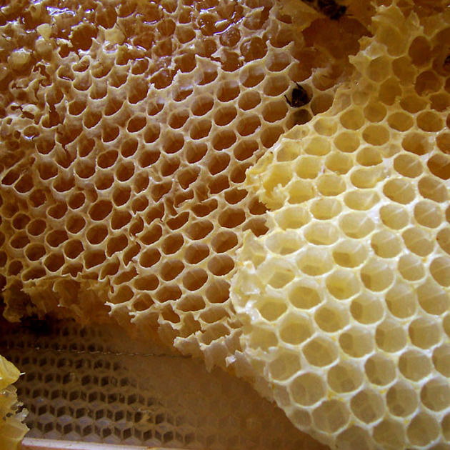
Honeycombing (lungs)
Citation, DOI, disclosures and article data
At the time the article was created Behrang Amini had no recorded disclosures.
View Behrang Amini's current disclosuresAt the time the article was last revised Henry Knipe had the following disclosures:
- Micro-X Ltd, Shareholder (past)
These were assessed during peer review and were determined to not be relevant to the changes that were made.
View Henry Knipe's current disclosures- Honeycomb lung
- Honeycombed lung
- Honeycomb lungs
- Honeycombed lungs
- Honey combing
Honeycombing describes small adjacent subpleural cystic structures in the context of established pulmonary fibrosis with destruction of lung parenchyma and loss of architecture 11. A single layer of cysts is now considered sufficient to apply this descriptor if other signs of pulmonary fibrosis are present11.
On this page:
Terminology
The terms microcystic honeycombing and macrocystic honeycombing are sometimes used ref. Exuberant honeycombing is a feature of connective tissue disease ref.
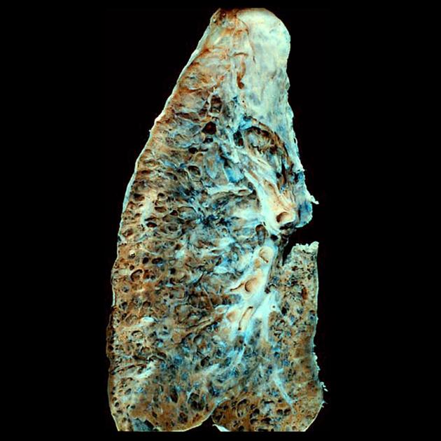
Pathology
The typical histological correlate of honeycombing is advanced traction bronchiolectasis surrounded by alveolar collapse 9. Microscopic honeycombing does not necessarily correlate with CT honeycombing 11. Pathologists regard honeycomb cysts as collapsed cystic secondary lobules lined by respiratory epithelium 12.
Radiographic features
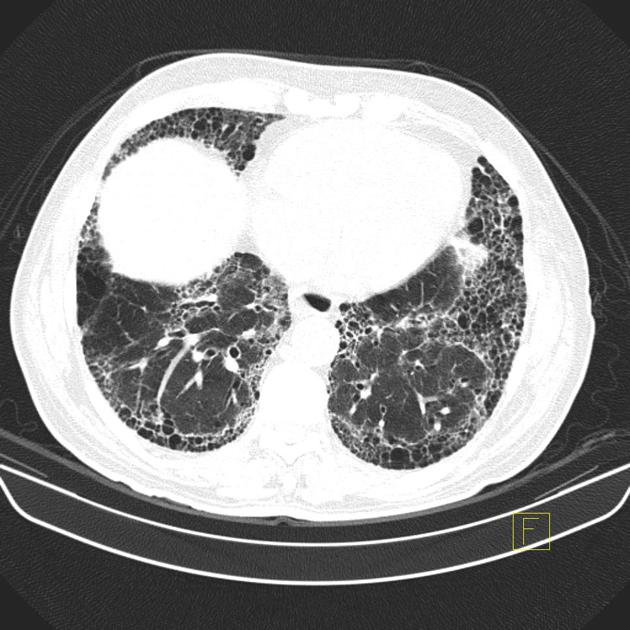
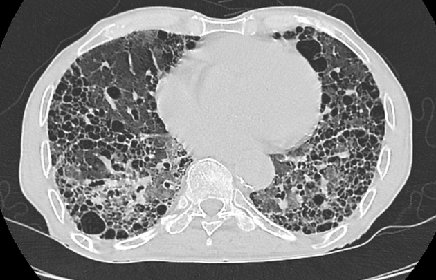
CT
Honeycombing and traction bronchiolectasis can be difficult to distinguish, but it is not surprising since traction bronchiolectasis is thought to progress to honeycombing. Honeycomb cysts are usually similar in size, <5 mm (range 3-25 mm), clustered in a row or stacked and with shared walls. The walls are relatively thick, around 1-3 mm. They are first seen adjacent to the pleura, usually in the basal lower lobes in areas of dense fibrosis 10.
History and etymology
The term “honeycomb lung” is thought to have originated in the 19th century in Germany and is thought to have first appeared in 1949 in a study by N Oswald and T Parkinson 5.
Differential diagnosis
-
in some situations
paraseptal emphysema (single layer of larger thin-walled juxta-pleural cyst-like spaces in non-fibrotic lung with upper lobe predominance) 10
airspace enlargement with fibrosis/smoking-related interstitial fibrosis (larger multi-layered or confluent thin-walled cysts, or cysts separated by normal lung which often spare the subpleural lung and lower zones) 10
airspace consolidation in the presence of pulmonary emphysema 8
Practical points
using the term "honeycombing" implies substantial pulmonary fibrosis is present, so it should be used with care 11
differentiation of honeycombing from emphysematous spaces with peripheral fibrosis can be difficult and the two conditions may coexist; the distribution is helpful, honeycombing is posterior lower zone predominant whereas emphysema with fibrosis is upper zone predominant 12
pathologists may find the diagnosis of honeycombing difficult as it can be difficult to distinguish from traction bronchiectasis; a clear histological definition would facilitate imaging evaluation 12
Quiz questions
References
- 1. Austin J, Müller N, Friedman P et al. Glossary of Terms for CT of the Lungs: Recommendations of the Nomenclature Committee of the Fleischner Society. Radiology. 1996;200(2):327-31. doi:10.1148/radiology.200.2.8685321 - Pubmed
- 2. Mueller-Mang C, Grosse C, Schmid K, Stiebellehner L, Bankier A. What Every Radiologist Should Know About Idiopathic Interstitial Pneumonias. Radiographics. 2007;27(3):595-615. doi:10.1148/rg.273065130 - Pubmed
- 3. Arakawa H & Honma K. Honeycomb Lung: History and Current Concepts. AJR Am J Roentgenol. 2011;196(4):773-82. doi:10.2214/AJR.10.4873 - Pubmed
- 4. Watadani T, Sakai F, Johkoh T et al. Interobserver Variability in the CT Assessment of Honeycombing in the Lungs. Radiology. 2013;266(3):936-44. doi:10.1148/radiol.12112516 - Pubmed
- 5. Johkoh T, Sakai F, Noma S et al. Honeycombing on CT; Its Definition, Pathologic Correlation, and Future Direction of Its Diagnosis. Eur J Radiol. 2014;83(1):27-31. doi:10.1016/j.ejrad.2013.05.012 - Pubmed
- 6. Wuyts W, Cavazza A, Rossi G, Bonella F, Sverzellati N, Spagnolo P. Differential Diagnosis of Usual Interstitial Pneumonia: When is It Truly Idiopathic? Eur Respir Rev. 2014;23(133):308-19. doi:10.1183/09059180.00004914 - Pubmed
- 7. Nishino M, Itoh H, Hatabu H. A Practical Approach to High-Resolution CT of Diffuse Lung Disease. Eur J Radiol. 2014;83(1):6-19. doi:10.1016/j.ejrad.2012.12.028 - Pubmed
- 8. Baik J, Ko J, Park H. Pitfalls in Radiographic Interpretation of Emphysema Patients. Can Assoc Radiol J. 2016;67(3):277-83. doi:10.1016/j.carj.2015.09.015 - Pubmed
- 9. Staats P, Kligerman S, Todd N, Tavora F, Xu L, Burke A. A Comparative Study of Honeycombing on High Resolution Computed Tomography with Histologic Lung Remodeling in Explants with Usual Interstitial Pneumonia. Pathology - Research and Practice. 2015;211(1):55-61. doi:10.1016/j.prp.2014.08.013 - Pubmed
- 10. Chae K, Hwang H, Duarte Achcar R et al. Central Role of CT in Management of Pulmonary Fibrosis. Radiographics. 2024;44(6):e230165. doi:10.1148/rg.230165 - Pubmed
- 11. Bankier A, MacMahon H, Colby T et al. Fleischner Society: Glossary of Terms for Thoracic Imaging. Radiology. 2024;310(2). doi:10.1148/radiol.232558
- 12. Galvin J & Franks T. Diffuse Parenchymal Lung Disease and the Inherent Limitations of a Glossary. Radiology. 2024;311(3):e240948. doi:10.1148/radiol.240948 - Pubmed
Incoming Links
- Chest curriculum
- Animal and animal produce inspired signs
- Interstitial lung pattern (radiograph)
- Diffuse alveolar damage
- Cystic lung disease
- Desquamative interstitial pneumonia
- Usual interstitial pneumonia
- Fibrotic non-specific interstitial pneumonitis
- IgG4-related lung disease
- Pneumothorax
- Idiopathic interstitial pneumonias (an approach on HRCT chest)
- Macrocystic honeycombing
- Pulmonary Langerhans cell histiocytosis
- Hard metal pneumoconiosis
- Asbestosis
- Bird fancier lung
- Microcystic honeycombing
- Pleuroparenchymal fibroelastosis
- Pulmonary cyst
- Food inspired signs
- Fibrotic hypersensitivity pneumonitis
- Cardiac arrest (CT)
- Methotrexate-induced pulmonary toxicity
- Rheumatoid interstitial lung disease (moderate)
- Usual interstitial pneumonia with concomitant intracavitary and endobronchial mycetomas
- Scleroderma-related interstitial lung disease
- Pulmonary fibrosis with unilateral lung transplant
- Usual interstitial pneumonia
- Polymyositis - with interstitial pneumonitis
- Chronic hypersensitivity pneumonitis
- Chronic hypersensitivity pneumonitis
- Usual interstitial pneumonia (probable)
- Honeycomb (photo)
- Honeycombing - pulmonary fibrosis
- Idiopathic pulmonary fibrosis
- Pulmonary fibrosis - UIP pattern
- Combined pulmonary fibrosis and emphysema - probable
- Usual interstitial pneumonia
- Non-specific interstitial pneumonia
- Combined UIP and emphysema in smoker
Related articles: Inspired signs
-
inanimate object inspired[+][+]
- accordion sign
- astronomical inspired
- ball of wool sign
- ball on tee sign (renal papillary necrosis)
- boot-shaped heart
- bowler hat sign
- bow tie sign
- box-shaped heart
- bucket handle appearance (disambiguation)
- chain of lakes sign
- champagne glass pelvis
- cobblestone appearance
- Coca-Cola bottle sign
- cockade sign (disambiguation)
- coin lesion
- collar button ulcer
- comb sign
- corduroy artifact
- corduroy sign
-
corkscrew sign (disambiguation)
- corkscrew sign (diffuse esophageal spasm)
- corkscrew sign (inner ear)
- corkscrew sign (midgut volvulus)
- crazy paving sign
- cupola sign
- curtain sign (lung ultrasound)
- dinner fork deformity
- dripping candle wax sign
- finger in glove sign
- fishhook ureters
- flame-shaped breast (gynecomastia)
- football sign (pneumoperitoneum)
- frozen pelvis
- ghost triad (gallbladder)
- ghost vertebra
- goblet sign
- ground glass opacity
- hockey stick sign (disambiguation)
- horseshoe (disambiguation)
- hourglass sign
- hurricane sign (cardiac SPECT)
- jail bar sign
- keyhole sign (disambiguation)
- leather bottle stomach
- light bulb sign (disambiguation)
- Lincoln log vertebra
- Mercedes-Benz sign (disambiguation)
- misty mesentery sign
- mosaic appearance (disambiguation)
- napkin ring sign
- open book fracture
- pearl necklace sign
- pencil in a cup
- picture frame vertebral body
- polka-dot sign
- rachitic rosary
- ribbon rib deformity
- ring shadow
- rugger jersey spine
- sack of marbles sign
- sail sign (disambiguation)
- scalpel sign
- spilled teacup sign
- stepladder sign (disambiguation)
-
string of pearls sign (disambiguation)
- string of pearls sign (abdominal radiograph of small bowel)
- string of pearls sign (polycystic ovarian syndrome)
- string of pearls sign (fibromuscular dysplasia)
- string of pearls sign (watershed infarction)
- Tam o' Shanter sign
- telephone receiver deformity
- thimble bladder
- tombstone iliac wings
- Venetian blind sign
- Venus necklace sign
- water bottle sign
-
weapon and munition inspired signs
- arrowhead sign
- bayonet artifact
- bayonet deformity
- boomerang sign (disambiguation)
- bullet-shaped vertebra
- cannonball metastases
- Cupid bow contour
- dagger sign
- double barrel sign
- halberd pelvis
- hatchet sign
- panzerherz
- pistol grip deformity
- saber-sheath trachea
- scimitar syndrome
-
target sign (disambiguation)
- double target sign (hepatic abscess)
- eccentric target sign (cerebral toxoplasmosis)
- reverse target sign (cirrhotic nodules)
- target sign (cholangiocarcinoma)
- target sign (choledocholithiasis)
- target sign (hepatic metastases)
- target sign (intussusception)
- target sign (neurofibromas)
- target sign (pyloric stenosis)
- target sign (tuberculosis)
- trident appearance
- Viking helmet sign
- white pyramid sign
- windswept knees
- wine bottle sign
-
vegetable and plant inspired[+][+]
- aubergine sign
- bamboo spine
- blade of grass sign
- celery stalk appearance (disambiguation)
- coconut left atrium
- coffee bean sign
- cotton wool appearance
- drooping lily sign
- ginkgo leaf sign (disambiguation)
- holly leaf sign
- iris sign
- ivy sign
- miliary opacities
- mistletoe sign
- onion signs (disambiguation)
- pine cone bladder
-
popcorn calcification (disambiguation)
- popcorn calcification (breast)
- popcorn calcification (chondroid lesions)
- popcorn calcification (fibrous dysplasia)
- popcorn calcification (osteogenesis imperfecta)
- popcorn calcification (pulmonary hamartomas)
- popcorn calcification (uterine fibroid)
- potato nodes
- rice signs (disambiguation)
- salt and pepper sign (disambiguation)
- tombstone iliac wings
- tree-in-bud
- tulip sign
- water lily sign
-
fruit inspired[+][+]
- apple core sign (disambiguation)
- apple-peel intestinal atresia
- banana and egg sign
- banana fracture
- banana sign
- berry aneurysm
- bowl of grapes sign
-
bunch of grapes sign (disambiguation)
- bunch of grapes sign (hydatidiform mole)
- bunch of grapes sign (bronchiectasis)
- bunch of grapes sign (IPMN)
- bunch of grapes sign (botryoid rhabdomyosarcoma)
- bunch of grapes sign (intracranial tuberculoma)
- bunch of grapes sign (intraosseous hemangiomas)
- bunch of grapes sign (multicystic dysplastic kidney)
- cashew nut sign
- lemon sign
- pear-shaped bladder
- strawberry gallbladder
- strawberry skull
- watermelon skin sign
-
animal and animal produce inspired[+][+]
- human
- mammals
- anteater nose sign
- antler sign
- batwing opacities
- bear paw sign
- beaver tail liver
- Brahma bull sign
- buffalo chest
- bull's eye sign (disambiguation)
- bunny waveform sign
- claw sign
- dog ear sign
- dog leg sign
- dromedary hump
- ears of the lynx sign
- eye of tiger sign
- feline esophagus
- giraffe pattern
- hidebound sign
- ivory phalanx
- ivory vertebra sign
- joint mouse
- leaping dolphin sign
- leopard skin sign
- moose head appearance
- panda sign
- piglet sign
- pleural mouse
- raccoon eyes sign
- rat bite erosions
- rat-tail sign
- Scottie dog sign
- Snoopy sign
- stag's antler sign
- staghorn calculus
- tiger stripe sign
-
zebra sign (disambiguation)
- zebra sign: (cerebellar hemorrhage)
- zebra spleen: arterial phase (spleen)
- zebra stripe sign (osteogenesis imperfecta)
- amphibians
- birds
- bird beak sign (disambiguation)
- bird's nest sign (lung)
- crow feet sign
- egg on a string sign
- eggshell calcification (breast)
- eggshell calcification (lymph nodes)
- gooseneck sign (endocardial cushion defect)
- gull wing appearance
- hummingbird sign
- owl eyes sign
- pooping duck sign
- sitting duck appearance
- swallowtail sign
- swan neck deformity
- winking owl sign
- fish and marine life
- reptiles
- arthropods
- micro-organisms
- fictional creatures
-
food inspired
- Cheerio sign (disambiguation)[+][+]
- chocolate cyst
- cottage loaf sign
- double Oreo cookie (glenoid labrum)
- doughnut sign (disambiguation)[+][+]
- hamburger sign (spine)
- head cheese sign (lungs)
- honeycombing (lungs)
- hot cross bun sign (pons)
- ice cream cone sign (middle ear ossicles)
- ice cream cone sign (vestibular schwannoma)
- licked candy stick appearance (bones)
- linguine sign (breast implants)
- macaroni sign
- omental cake
- Oreo cookie (heart)
- pancake organ (disambiguation)[+][+]
- Polo mint sign
- salad oil sign (breast implants)
- sandwich sign (disambiguation)[+][+]
- sandwich vertebra
- sausage digit
- spaghetti sign
- Swiss cheese sign
-
alphabet inspired[+][+]
- A line (US artifact)
- C sign (MSK)
- delta sign (disambiguation)
- E sign
- H-shaped vertebra
- H sign
- J-shaped sella
- J sign (shoulder)
- L sign (brain)
- lambda sign (disambiguation)
- M sign
- omega epiglottis
- O sign (gastric banding)
- P sign (epiglottis)
- S sign of Golden
- tau sign
- T sign (disambiguation)
- U fibers
- U-figure (pelvis)
- U sign (brain)
- V sign (disambiguation)
- W hernia
- X-marks-the-spot sign
- Y sign (epidural lipomatosis)
- Z deformity
-
Christmas inspired[+][+]
- Christmas tree bladder in neurogenic bladder
- holly leaf sign in calcified pleural plaques
- ivy sign in leptomeningeal enhancement
- nutcracker esophagus in esophageal dysmotility
- shepherd's crook deformity of the femur in fibrous dysplasia
- snowcap sign in avascular necrosis
- snowman sign (disambiguation)
- snowstorm appearance in complete hydatidiform mole and testicular microlithiasis
- miscellaneous[+][+]


 Unable to process the form. Check for errors and try again.