
Accessory ossicles of the foot
Citation, DOI, disclosures and article data
Citation:
Bashir U, Niknejad M, Rasuli B, et al. Accessory ossicles of the foot. Reference article, Radiopaedia.org (Accessed on 21 Mar 2025) https://doi.org/10.53347/rID-18879
rID:
18879
Article created:
26 Jul 2012,
Usman Bashir
Disclosures:
At the time the article was created Usman Bashir had no recorded disclosures.
View Usman Bashir's current disclosures
Last revised:
Disclosures:
At the time the article was last revised Mohammad Taghi Niknejad had no financial relationships to ineligible companies to disclose.
View Mohammad Taghi Niknejad's current disclosures
Revisions:
27 times, by
10 contributors -
see full revision history and disclosures
Systems:
Sections:
Tags:
Synonyms:
- Secondary ossification centers of the foot
- Accessory ossicles of feet
- Accessory ossicles of foot
- Accessory ossicles of the feet
- Accessory tarsal bones
- Accessory tarsal ossicles
- Supernumerary tarsal bone
- Supernumerary ossicles of foot
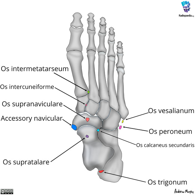
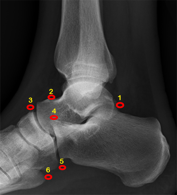
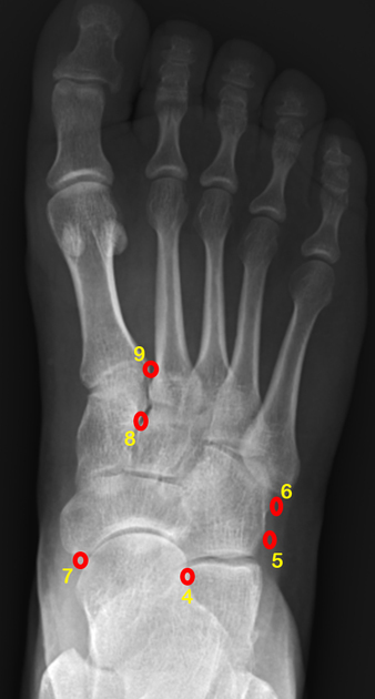
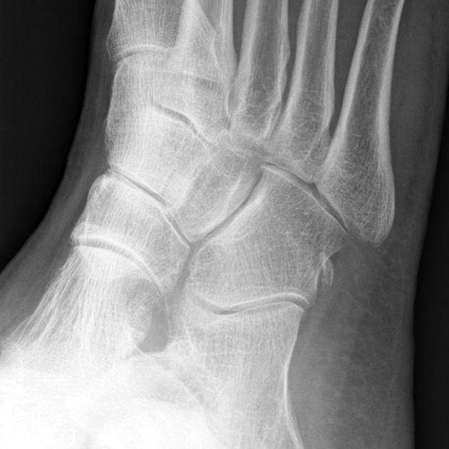
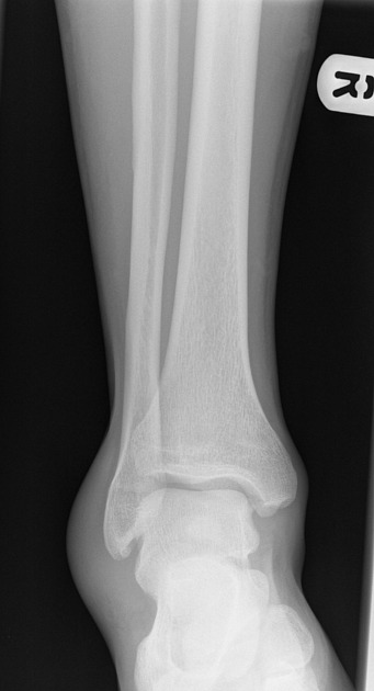
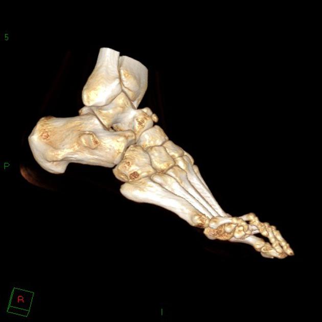
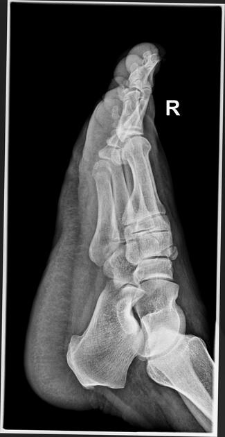
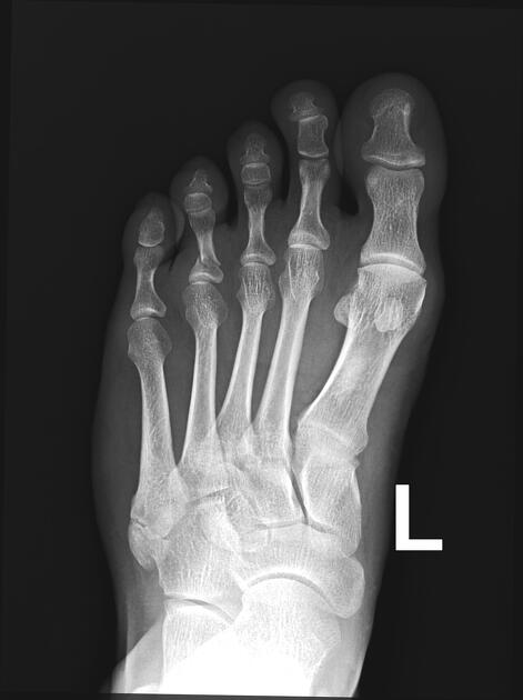
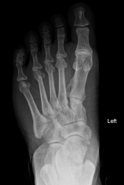
Accessory ossicles of the feet are common developmental variants with almost 40 having been described. Some of the more common include 1-4:
- os peroneum
- os subfibulare
- os subtibiale
- os tibiale externum (accessory navicular)
- os trigonum
- os calcaneus secundaris
- os calcanei accessorium 6
- os intermetatarseum
- pars peronea metatarsalis primi (pars peronea metatarsalia)
- os supratalare
- bipartite hallux sesamoid
- os supranaviculare
- os infranaviculare (cuneonavicular ossicle) 5
- os intercuneiforme
- os vesalianum pedis
- os sustentaculi
- os subcalcis
- os cuboideum secundarium
- os talotibiale
- os paracuneiforme
- os aponeurosis plantaris
- os cuneometatarsale plantare
- os talus secundarius
- os cuneometatarsale I tibiale
- os cuneo–I metatarsale-II dorsale
Knowledge of their presence is helpful so that they are not misdiagnosed as fractures.

References
- 1. Theodore Eliot Keats, Mark W. Anderson. Atlas of Normal Roentgen Variants That May Simulate Disease. (2007) ISBN: 9780323043007 - Google Books
- 2. Coskun N, Yuksel M, Cevener M et al. Incidence of Accessory Ossicles and Sesamoid Bones in the Feet: A Radiographic Study of the Turkish Subjects. Surg Radiol Anat. 2009;31(1):19-24. doi:10.1007/s00276-008-0383-9 - Pubmed
- 3. Kalantari BN, Seeger LL, Motamedi K, Chow K. Accessory ossicles and sesamoid bones: Spectrum of pathology and imaging evaluation. Appl Radiol. 2007;36(10):28-37. Appl Radiol (full text)
- 4. Thomas H. Berquist. Imaging of the Foot and Ankle. (2011) ISBN: 9781605475721 - Google Books
- 5. Robert Christman. Foot and Ankle Radiology. (2014) ISBN: 9781496304391 - Google Books
- 6. Keles-Celik N, Kose O, Sekerci R, Aytac G, Turan A, Güler F. Accessory Ossicles of the Foot and Ankle: Disorders and a Review of the Literature. Cureus. 2017;9(11):e1881. doi:10.7759/cureus.1881 - Pubmed
Incoming Links
Articles:
- Bipartite medial cuneiform
- Os intercuneiforme
- Ankle radiograph (an approach)
- Os supranaviculare
- Os vesalianum pedis
- Foot radiograph (checklist)
- Os talotibiale
- Accessory ossicles
- Fibula
- Os subtibiale
- Os sustentaculi
- Os intermetatarseum
- Pars peronea metatarsalis primi
- Accessory ossicles of the lower limb
- Os cuboideum secundarium
Cases:
- Calcaneal fracture
- Osteoid osteoma of the medial cuneiform
- Os supranaviculare - symptomatic
- Os supranaviculare
- Os intermetatarseum and os calcaneus secundarius
- Os vesalianum pedis
- Polymetatarsia without polydactyly
- Os subtibiale
- Accessory ossicles of the foot (illustration)
- Accessory bones of the foot - os tibiale externum and os peroneum
- Unfused secondary ossification centre of the medial malleolus
- Os intermetatarseum
- Os subfibulare
- Interphalangeal joint sesamoid of great toe
- Os calcaneus secundarius
- Os supratalare
- Os subtibiale
- Exostosis of the talus
- Os calcaneus secundarius
- Os infranaviculare
Multiple choice questions:
Related articles: Anatomy: Lower limb
- skeleton of the lower limb
- joints of the lower limb
-
hip joint
- ligaments
- muscles
- additional structures
- hip joint capsule
- zona orbicularis
- iliotibial band
-
hip bursae
- anterior
- iliopsoas bursa (iliopectineal bursa)
- lateral
- subgluteal bursae
- greater trochanteric bursa (subgluteus maximus bursa)
- subgluteus medius bursa
- subgluteus minimus bursa
- gluteofemoral bursa
- subgluteal bursae
- postero-inferior
- anterior
- ossification centers
-
knee joint
- ligaments
- anterior cruciate ligament
- posterior cruciate ligament
- medial collateral ligament
- lateral collateral ligament
- meniscofemoral ligament (mnemonic)
-
posterolateral ligamentous complex
- arcuate ligament
- patellar tendon and quadriceps tendon
- anterolateral ligament
- posterior oblique ligament
- oblique popliteal ligament
- medial patellofemoral ligament
- additional structures
- extensor mechanism of the knee
- groove for the popliteus tendon
- knee bursae
- anterior bursae
- medial bursae
- lateral bursae
- posterior bursae
- knee capsule
- lateral patellar retinaculum
- medial patellar retinaculum
- menisci
- pes anserinus (mnemonic)
- ossification centers
- ligaments
- tibiofibular joints
-
ankle joint
- regional anatomy
- medial ankle
- lateral ankle
- anterior ankle
- ligaments
- medial collateral (deltoid) ligament
- lateral collateral ligament
- additional structures
- ankle bursae
- ossification centers of the ankle
- variants
- regional anatomy
- foot joints
- subtalar joint
- mid-tarsal (Chopart) joint
-
tarsometatarsal (Lisfranc) joint
- ligaments
- intermetatarsal joint
- metatarsophalangeal joint
- interphalangeal joint
- ossification centers
-
hip joint
- spaces of the lower limb
-
muscles of the lower limb
- muscles of the pelvic group
- muscles of the thigh
- muscles of the leg
- anterior compartment of the leg
- posterior compartments of the leg
- lateral compartment of the leg
- muscles of the foot
- dorsal muscles
- plantar muscles
- 1st layer
- 2nd layer
- 3rd layer
- 4th layer
- accessory muscles of the lower limb
- accessory gluteal muscles
-
accessory muscles of the ankle
- accessory peroneal muscles
- accessory flexor digitorum longus muscle
- accessory soleus muscle
- peroneocalcaneus internus muscle
- tibiocalcaneus internus muscle
- extensor hallucis capsularis tendon
- anterior fibulocalcaneus muscle
- accessory extensor digiti secundus muscle
- tibioastragalus anticus of Gruber muscle
- vascular supply of the lower limb
- arterial supply of the lower limb
- venous drainage of the lower limb
- innervation of the lower limb
- lymphatic system of the lower limb
- lymphatic pathways
- anteromedial group
- anterolateral group
- posteromedial group
- posterolateral group
- lower limb lymph nodes
- lymphatic pathways


 Unable to process the form. Check for errors and try again.