
The anal canal is the terminal part of the gastrointestinal tract, whilst the anus (plural: anuses or ani) specifically refers to the opening separating the anal canal from the outside, at the distal most aspect of the anal verge. Anatomically, the anal canal is referred to as the terminal alimentary tract between the dentate line and anal verge. However, histologically it extends more proximally and includes the anal columns (of Morgagni) and anal sinuses. Surgically, the anal canal is referred to as the portion of bowel between the anorectal sling and the anal verge.
The anal margin is arbitrarily defined as the 5 cm of skin (radius) surrounding the anal verge.
On this page:
Gross anatomy
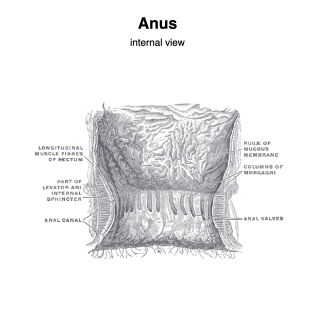
The anal canal measures ~4 cm long and is continuous with the rectum at the anorectal junction, which is the angle (the anorectal angle) the rectum makes at the levator ani.
The anal canal is a muscular tube, like the rest of the gastrointestinal tract, with the muscular layer forming the external and internal anal sphincters.
The anal canal contains longitudinal folds of mucosa, termed the anal columns which join inferiorly to form semicircular anal valves. The anal valves collectively form the dentate (pectinate) line, which marks the embryological anal membrane and divides the anal canal into an upper and lower parts, differing in structure and neurovascular supply.
The dentate line is a "watershed area" and the exact transition of epithelium and neurovascular supply is variable:
above the dentate line, the epithelium is mucous membrane (columnar epithelium) like the rest of the gastrointestinal tract
below the dentate line is a transition zone, lined by nonkeratinised stratified squamous epithelium known as the anal pecten 3
further inferiorly the anal pecten ends at the anocutaneous (white) line, where the epithelium becomes true skin (i.e. keratinized stratified squamous with hair and sebaceous glands) 3
Arterial supply
above dentate line: superior rectal artery (from inferior mesenteric artery); small contributions from middle rectal artery (directly from internal iliac artery) and median sacral arteries
below dentate line: inferior rectal artery (from internal pudendal artery)
Venous drainage
Venous drainage richly anastomoses with the rectal venous plexus
above dentate line: superior rectal vein to inferior mesenteric vein (portal venous system)
below dentate line: inferior and middle rectal veins to internal pudendal vein, a tributary of the internal iliac vein (systemic venous system)
The anal canal is a site of portosystemic anastomosis.
Lymphatic drainage
above dentate line: internal iliac nodes
below dentate line: superficial inguinal nodes
Innervation
-
above dentate line and internal anal sphincter
parasympathetic and afferent sensory: pelvic splanchnic nerves
-
below dentate line and external anal sphincter
Development
The upper part of the anal canal derives from the dorsal compartment of the cloaca (endoderm) and the lower part is derived from proctodeum (ectoderm).
Related conditions
-
1 in 1500-5000 newborns
failure of the anus to correctly form
may vary from stenosis to blind anal canal/rectum to absent anal canal


 Unable to process the form. Check for errors and try again.