
Bony pelvis
Citation, DOI, disclosures and article data
At the time the article was created Jeremy Jones had no recorded disclosures.
View Jeremy Jones's current disclosuresAt the time the article was last revised Tariq Walizai had no financial relationships to ineligible companies to disclose.
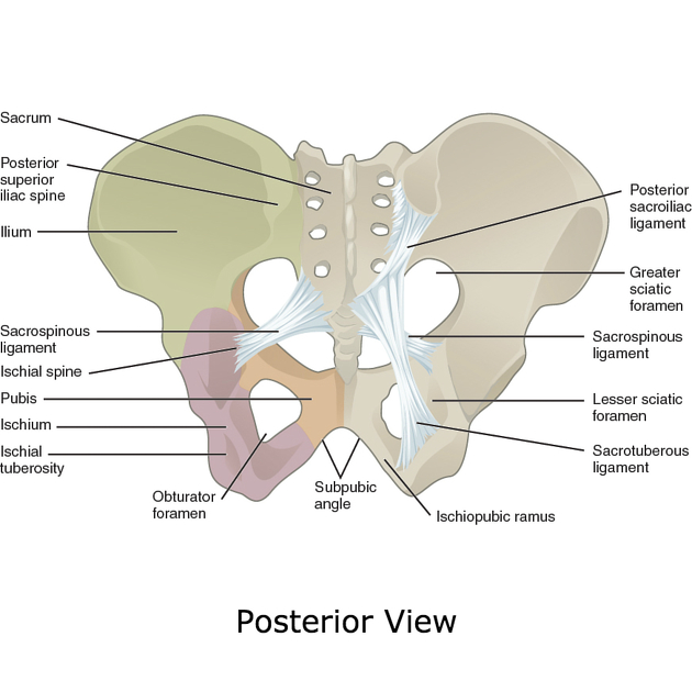
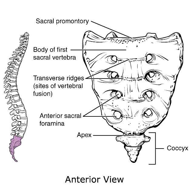
View Tariq Walizai's current disclosuresThe bony pelvis is formed by the sacrum and coccyx and a pair of hip bones (os coxae or innominate bones), comprising the ischium, pubis and ilium and are part of the appendicular skeleton.
Its primary function is the transmission of forces from the axial skeleton to the lower limbs as well as supporting the pelvic viscera.
Gross anatomy
In childhood, each hip bone consists of three separate bones (ilium, ischium and pubis) connected by the triradiate cartilage. Around puberty, these bones fuse.
The two hip bones are joined anteriorly at the symphysis pubis and posteriorly to the sacrum at the sacroiliac joints. The pelvic bones incorporate the acetabulum, which articulates with the proximal femur at the hip joint.
Sex differences
Differences between the males and female bony pelvis arise as an adaptation of the female pelvis to childbearing 3:
-
pelvic inlet shape
round or oval in females
heart-shaped in males
infrapubic angle is greater than 90° in females
greater sciatic notch is wider in females
acetabulum faces more anteriorly in females
sacrum more triangular and shorter in females
oval or triangular obturator foramen in females
The shape of the female bony pelvis can be described using the following terms 3:
gynaecoid pelvis (50%): normal female type
anthropoid pelvis (25%): long AP diameters, short transverse diameters and narrow infrapubic angle
android pelvis (20%): male type with conical shaped pelvic cavity and heart-shaped pelvic inlet
platypelloid ("flat female") pelvis (5%): short AP diameters, long transverse diameters and wide infrapubic angle
Pelvic apertures
The pelvic brim defines the pelvic inlet and the following structures contribute to it 2:
pubic crest
pectin pubis
arcuate line (of the ilium)
sacral ala
Pelvic outlet is formed by the following structures 2:
pubic arch
inferior margin of the pubic symphysis
pubic rami
ischial rami
sacrum and coccyx
References
- 1. Gray's Anatomy. Churchill Livingstone. (2011) ISBN:0443066841. Read it at Google Books - Find it at Amazon
- 2. Rosse C, Gaddum-Rosse P, Hollinshead WH. Hollinshead's textbook of anatomy. Lippincott Williams & Wilkins. (1997) ISBN:0397512562. Read it at Google Books - Find it at Amazon
- 3. Butler P, Mitchell A, Healy JC. Applied Radiological Anatomy. (2012) ISBN:0521766664. Read it at Google Books - Find it at Amazon
Incoming Links
- Pelvic kidney
- Mandibular fracture
- Sacrospinous ligament
- Primary bone lymphoma
- Pelvic fractures
- Appendicular skeleton
- Prostate MRI (an approach)
- Innominate bones
- Pelvis
- Open book pelvic injury
- Sacrum
- Pelvic fractures (summary)
- Ilium vs ileum
- Faulty fetal packing
- Secondary involvement of the bone with lymphoma
- Sacrotuberous ligament
Related articles: Anatomy: Abdominopelvic
- skeleton of the abdomen and pelvis
- muscles of the abdomen and pelvis
- spaces of the abdomen and pelvis
- anterior abdominal wall
- posterior abdominal wall
- abdominal cavity
- pelvic cavity
- perineum
- abdominal and pelvic viscera
- gastrointestinal tract
- spleen
- hepatobiliary system
-
endocrine system
-
adrenal gland
- adrenal vessels
- chromaffin cells
- variants
- pancreas
- organs of Zuckerkandl
-
adrenal gland
-
urinary system
-
kidney
- renal pelvis
- renal sinus
- avascular plane of Brodel
-
variants
- number
- fusion
- location
- shape
- ureter
- urinary bladder
- urethra
- embryology
-
kidney
- male reproductive system
-
female reproductive system
- vulva
- vagina
- uterus
- adnexa
- Fallopian tubes
- ovaries
- broad ligament (mnemonic)
- variant anatomy
- embryology
- blood supply of the abdomen and pelvis
- arteries
-
abdominal aorta
- inferior phrenic artery
- celiac artery
- superior mesenteric artery
- middle suprarenal artery
- renal artery (variant anatomy)
- gonadal artery (ovarian artery | testicular artery)
- inferior mesenteric artery
- lumbar arteries
- median sacral artery
-
common iliac artery
- external iliac artery
-
internal iliac artery (mnemonic)
- anterior division
- umbilical artery
- superior vesical artery
- obturator artery
- vaginal artery
- inferior vesical artery
- uterine artery
- middle rectal artery
-
internal pudendal artery
- inferior rectal artery
-
perineal artery
- posterior scrotal artery
- transverse perineal artery
- artery to the bulb
- deep artery of the penis/clitoris
- dorsal artery of the penis/clitoris
- inferior gluteal artery
- posterior division (mnemonic)
- variant anatomy
- anterior division
-
abdominal aorta
- portal venous system
- veins
- anastomoses
- arterioarterial anastomoses
- portal-systemic venous collateral pathways
- watershed areas
- arteries
- lymphatics
- innervation of the abdomen and pelvis
- thoracic splanchnic nerves
- lumbar plexus
-
sacral plexus
- lumbosacral trunk
- sciatic nerve
- superior gluteal nerve
- inferior gluteal nerve
- nerve to piriformis
- perforating cutaneous nerve
- posterior femoral cutaneous nerve
- parasympathetic pelvic splanchnic nerves
- pudendal nerve
- nerve to quadratus femoris and inferior gemellus muscles
- nerve to internal obturator and superior gemellus muscles
- autonomic ganglia and plexuses


 Unable to process the form. Check for errors and try again.