
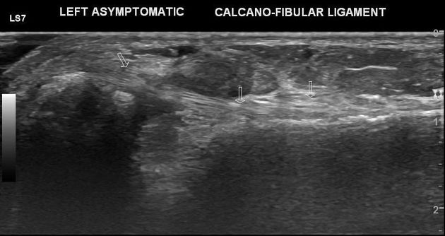
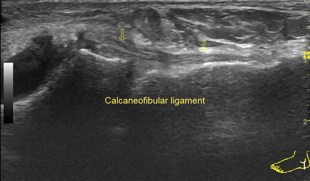
Calcaneofibular ligament
Citation, DOI, disclosures and article data
At the time the article was created Henry Knipe had no recorded disclosures.
View Henry Knipe's current disclosuresAt the time the article was last revised Yahya Baba had no financial relationships to ineligible companies to disclose.
View Yahya Baba's current disclosures- Calcaneo-fibular ligament
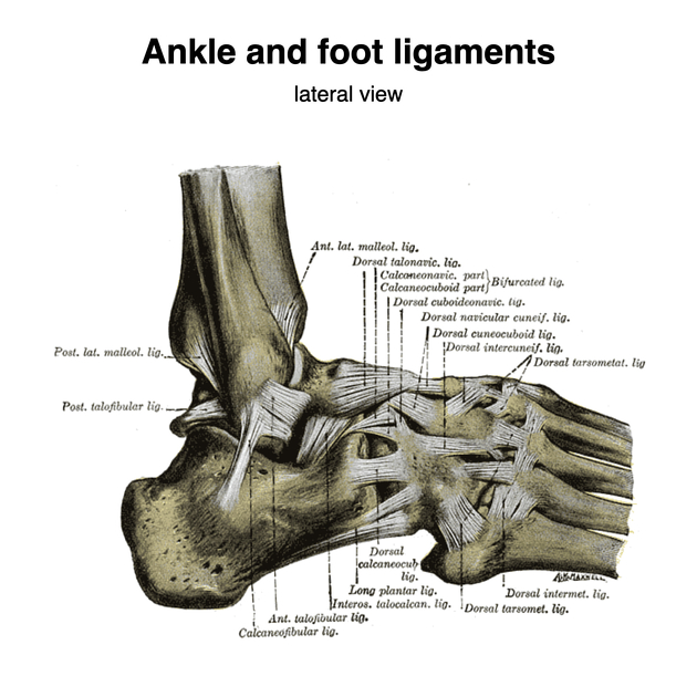
The calcaneofibular ligament (CFL) is the middle ligament of the lateral collateral ligament complex of the ankle and stabilizes both the ankle and subtalar joints.
On this page:
Gross anatomy
The CFL is an extracapsular round cord measuring 20-25 mm long x 6-8 mm width. Its origin is distal to the anterior talofibular ligament on the tip of the lateral malleolus and courses posteroinferiorly and medially to insert onto the lateral surface of the calcaneus.
Relations
lies deep to the peroneal tendons 2
Radiographic appearance
MRI
often only partially imaged on coronal and axial images
PD: uniformly low signal 1
Related pathology
References
- 1. Perrich K, Goodwin D, Hecht P, Cheung Y. Ankle Ligaments on MRI: Appearance of Normal and Injured Ligaments. AJR Am J Roentgenol. 2009;193(3):687-95. doi:10.2214/AJR.08.2286 - Pubmed
- 2. Alan S. Banks (Editor), Michael S. Downey (Editor). McGlamry's Comprehensive Textbook of Foot and Ankle Surgery. (2001) ISBN: 9780683304718 - Google Books
- 3. Meir Nyska, Gideon Mann. The Unstable Ankle. (2002) ISBN: 9780880118026 - Google Books
Incoming Links
- Ankle radiograph (an approach)
- Anterolateral recess of the ankle joint
- Chronic ankle instability
- Ankle joint
- Medical abbreviations and acronyms (C)
- Anterior talofibular ligament injury
- Lateral ankle sprain
- MRI of the ankle (an approach)
- Posterior talofibular ligament
- Sural neuropathy
- Calcaneofibular ligament injury
- Fibula
- Ankle protocol (MRI)
- Subtalar instability
- Lateral collateral ligament of the ankle
- Anterior talofibular ligament bony avulsion - pediatric
- Lateral malleolus avulsion fracture
- Peroneus brevis tear
- Calcaneofibular ligament bony avulsion
- Peroneus quartus muscle
- Anterior talofibular ligament injury with variant anatomy
- Anterior talofibular ligament bony avulsion - paediatric
- Midtarsal sprain - ultrasound
- Anterior inferior tibiofibular ligament injury
- Anterior talofibular ligament bony avulsion - paediatric
- Wagstaffe-Le Fort fracture
- Ankle extensor retinaculum and lateral ligaments injuries - ultrasound
- Anterior talofibular ligament bony avulsion - paediatric
- Ankle ligaments injury
- Anterior inferior tibiofibular ligament injury
- Lateral ankle ligaments injury
- Lateral talocrural ligaments (Gray's illustration)
- Ankle and foot ligaments (Gray's illustration)
- Anterior talofibular ligament bony avulsion from fibula
- Fibroma (ankle joint)
Related articles: Anatomy: Lower limb
- skeleton of the lower limb
- joints of the lower limb
-
hip joint
- ligaments
- muscles
- additional structures
- hip joint capsule
- zona orbicularis
- iliotibial band
-
hip bursae
- anterior
- iliopsoas bursa (iliopectineal bursa)
- lateral
- subgluteal bursae
- greater trochanteric bursa (subgluteus maximus bursa)
- subgluteus medius bursa
- subgluteus minimus bursa
- gluteofemoral bursa
- subgluteal bursae
- postero-inferior
- anterior
- ossification centers
-
knee joint
- ligaments
- anterior cruciate ligament
- posterior cruciate ligament
- medial collateral ligament
- lateral collateral ligament
- meniscofemoral ligament (mnemonic)
-
posterolateral ligamentous complex
- arcuate ligament
- patellar tendon and quadriceps tendon
- anterolateral ligament
- posterior oblique ligament
- oblique popliteal ligament
- medial patellofemoral ligament
- additional structures
- extensor mechanism of the knee
- groove for the popliteus tendon
- knee bursae
- anterior bursae
- medial bursae
- lateral bursae
- posterior bursae
- knee capsule
- lateral patellar retinaculum
- medial patellar retinaculum
- menisci
- pes anserinus (mnemonic)
- ossification centers
- ligaments
- tibiofibular joints
-
ankle joint
- regional anatomy
- medial ankle
- lateral ankle
- anterior ankle
- ligaments
- medial collateral (deltoid) ligament
- lateral collateral ligament
- additional structures
- ankle bursae
- ossification centers of the ankle
- variants
- regional anatomy
- foot joints
- subtalar joint
- mid-tarsal (Chopart) joint
-
tarsometatarsal (Lisfranc) joint
- ligaments
- intermetatarsal joint
- metatarsophalangeal joint
- interphalangeal joint
- ossification centers
-
hip joint
- spaces of the lower limb
-
muscles of the lower limb
- muscles of the pelvic group
- muscles of the thigh
- muscles of the leg
- anterior compartment of the leg
- posterior compartments of the leg
- lateral compartment of the leg
- muscles of the foot
- dorsal muscles
- plantar muscles
- 1st layer
- 2nd layer
- 3rd layer
- 4th layer
- accessory muscles of the lower limb
- accessory gluteal muscles
-
accessory muscles of the ankle
- accessory peroneal muscles
- accessory flexor digitorum longus muscle
- accessory soleus muscle
- peroneocalcaneus internus muscle
- tibiocalcaneus internus muscle
- extensor hallucis capsularis tendon
- anterior fibulocalcaneus muscle
- accessory extensor digiti secundus muscle
- tibioastragalus anticus of Gruber muscle
- vascular supply of the lower limb
- arterial supply of the lower limb
- venous drainage of the lower limb
- innervation of the lower limb
- lymphatic system of the lower limb
- lymphatic pathways
- anteromedial group
- anterolateral group
- posteromedial group
- posterolateral group
- lower limb lymph nodes
- lymphatic pathways


 Unable to process the form. Check for errors and try again.