
Infrapatellar fat pad
Citation, DOI, disclosures and article data
At the time the article was created Yuranga Weerakkody had no recorded disclosures.
View Yuranga Weerakkody's current disclosuresAt the time the article was last revised Alex Zheng had no financial relationships to ineligible companies to disclose.
View Alex Zheng's current disclosures- Hoffa fat pad
- Hoffa's fat pad
- Hoffa's fat pads
- Infrapatellar fat pads
- Hoffa fat pads
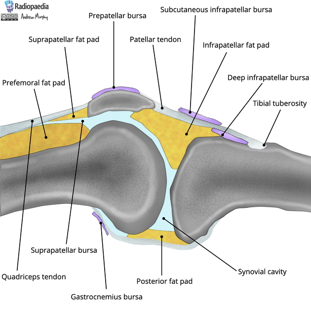
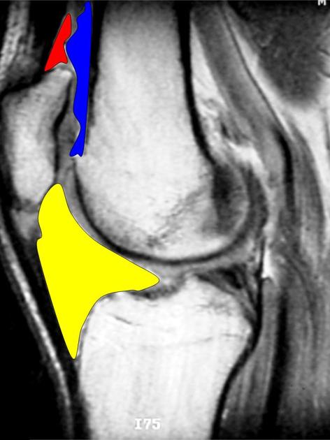
The infrapatellar fat pad, also known as Hoffa fat pad, is the largest of the anterior knee fat pads. It is located immediately posterior to the patellar tendon and is traversed during knee arthroscopy.
On this page:
Anatomy
Boundaries
-
anterior
anteroinferior: patellar tendon
anterosuperior: patella
-
posterior
posterocentral: knee joint (femorotibial articulation)
posteroinferior: tibia
posterosuperior: femur
History and etymology
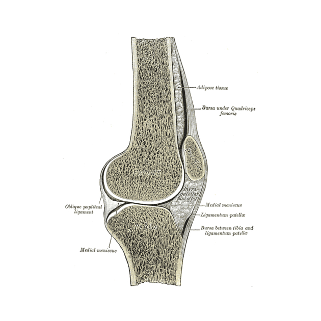
It was initially described by German surgeon Albert Hoffa (1859-1907) in 1904 3.
Related pathology
patellar tendon lateral femoral condyle friction syndrome (Hoffa fat pad impingement syndrome)
Quiz questions
References
- 1. Jacobson JA, Lenchik L, Ruhoy MK, Schweitzer ME, Resnick D. MR imaging of the infrapatellar fat pad of Hoffa. (1997) Radiographics : a review publication of the Radiological Society of North America, Inc. 17 (3): 675-91. doi:10.1148/radiographics.17.3.9153705 - Pubmed
- 2. Draghi F, Ferrozzi G, Urciuoli L, Bortolotto C, Bianchi S. Hoffa's fat pad abnormalities, knee pain and magnetic resonance imaging in daily practice. (2016) Insights into imaging. 7 (3): 373-83. doi:10.1007/s13244-016-0483-8 - Pubmed
- 3. Zeng N, Yan Z, Chen X, Ni G. Infrapatellar Fat Pad and Knee Osteoarthritis. Aging Dis. 2020;11(5):1317-28. doi:10.14336/AD.2019.1116 - Pubmed
Incoming Links
- MRI Osteoarthritis Knee Score (MOAKS)
- Sinding-Larsen-Johansson disease
- Anterior interval lesion
- Inferior medial genicular artery
- Osgood-Schlatter disease
- Hoffa fracture
- Patellar tendon
- Arthrofibrosis
- Ligamentum mucosum
- Hoffa fat pad ganglion cyst
- Prefemoral fat pad
- Inferior lateral genicular artery
- Hoffa fat pad herniation
- Knee joint
- Anterior knee fat pads
- Anterior cruciate ligament ganglion cyst
- Patellar tendon-lateral femoral condyle friction syndrome
- Patellar tendon rupture
- Soft tissue chondroma of the Hoffa fat pad
- MRI of the knee (an approach)
- Hoffa fat pad ganglion cyst
- Gout
- Anteromedial meniscofemoral ligament
- Intracapsular chondroma - knee
- Deep infrapatellar bursitis
- Hoffa's fat pad (Gray's illustration)
- Patellar chondral avulsion fracture
- Patellar dislocation with chondral injury
- Chondroblastoma - tibia
- Synovial haemangioma - knee
- Ligamentum mucosum (infrapatellar plica)
- Synovial haemangioma
- Intracapsular chondroma - knee
- Patellar tendon rupture
- Infrapatellar fat pad hemangioma
- Pigmented villonodular synovitis (PVNS) - focal
- Hoffa's fat pad ganglion cyst
- Intracapsular chondroma of the knee
- MRI knee - sagittal (anatomy quiz)
- Quadriceps fat pad impingement syndrome
Related articles: Anatomy: Lower limb
- skeleton of the lower limb
- joints of the lower limb
-
hip joint
- ligaments
- muscles
- additional structures
- hip joint capsule
- zona orbicularis
- iliotibial band
-
hip bursae
- anterior
- iliopsoas bursa (iliopectineal bursa)
- lateral
- subgluteal bursae
- greater trochanteric bursa (subgluteus maximus bursa)
- subgluteus medius bursa
- subgluteus minimus bursa
- gluteofemoral bursa
- subgluteal bursae
- postero-inferior
- anterior
- ossification centers
-
knee joint
- ligaments
- anterior cruciate ligament
- posterior cruciate ligament
- medial collateral ligament
- lateral collateral ligament
- meniscofemoral ligament (mnemonic)
-
posterolateral ligamentous complex
- arcuate ligament
- patellar tendon and quadriceps tendon
- anterolateral ligament
- posterior oblique ligament
- oblique popliteal ligament
- medial patellofemoral ligament
- additional structures
- extensor mechanism of the knee
- groove for the popliteus tendon
- knee bursae
- anterior bursae
- medial bursae
- lateral bursae
- posterior bursae
- knee capsule
- lateral patellar retinaculum
- medial patellar retinaculum
- menisci
- pes anserinus (mnemonic)
- ossification centers
- ligaments
- tibiofibular joints
-
ankle joint
- regional anatomy
- medial ankle
- lateral ankle
- anterior ankle
- ligaments
- medial collateral (deltoid) ligament
- lateral collateral ligament
- additional structures
- ankle bursae
- ossification centers of the ankle
- variants
- regional anatomy
- foot joints
- subtalar joint
- mid-tarsal (Chopart) joint
-
tarsometatarsal (Lisfranc) joint
- ligaments
- intermetatarsal joint
- metatarsophalangeal joint
- interphalangeal joint
- ossification centers
-
hip joint
- spaces of the lower limb
-
muscles of the lower limb
- muscles of the pelvic group
- muscles of the thigh
- muscles of the leg
- anterior compartment of the leg
- posterior compartments of the leg
- lateral compartment of the leg
- muscles of the foot
- dorsal muscles
- plantar muscles
- 1st layer
- 2nd layer
- 3rd layer
- 4th layer
- accessory muscles of the lower limb
- accessory gluteal muscles
-
accessory muscles of the ankle
- accessory peroneal muscles
- accessory flexor digitorum longus muscle
- accessory soleus muscle
- peroneocalcaneus internus muscle
- tibiocalcaneus internus muscle
- extensor hallucis capsularis tendon
- anterior fibulocalcaneus muscle
- accessory extensor digiti secundus muscle
- tibioastragalus anticus of Gruber muscle
- vascular supply of the lower limb
- arterial supply of the lower limb
- venous drainage of the lower limb
- innervation of the lower limb
- lymphatic system of the lower limb
- lymphatic pathways
- anteromedial group
- anterolateral group
- posteromedial group
- posterolateral group
- lower limb lymph nodes
- lymphatic pathways


 Unable to process the form. Check for errors and try again.