
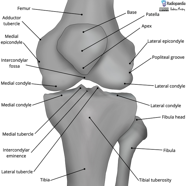
Intercondylar area
Citation, DOI, disclosures and article data
At the time the article was created Marlyson Olivier had no recorded disclosures.
View Marlyson Olivier's current disclosuresAt the time the article was last revised Alex Zheng had no financial relationships to ineligible companies to disclose.
View Alex Zheng's current disclosures- Intercondylar region
The intercondylar area is the rough, central part of the tibial plateau.
Gross anatomy
The intercondylar area is located between the proximal articular surfaces of the medial and lateral tibial condyles. It is non-articular. In the middle of the intercondylar area are:
intercondylar eminence: narrow, raised central part of the intercondylar area
medial and lateral intercondylar tubercles or spines: arise from the central aspect of the intercondylar eminence
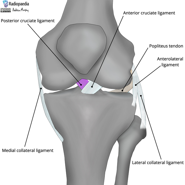
Attachments
Six facets are present for the attachment of the cruciate ligaments and menisci. A useful mnemonic for the six facets is 'MCLLMC', arranged from anterior to posterior:
medial meniscus anterior horn: at the anterior margin of the intercondylar area
anterior cruciate ligament: anteromedial to the anterior horn of the lateral meniscus attachment
lateral meniscus anterior horn: anterior to the medial intercondylar tubercle
lateral meniscus posterior horn: posterior to the lateral intercondylar tubercle
medial meniscus posterior horn: posterior to the attachment of the posterior horn of the lateral meniscus and anterior to the attachment of the posterior cruciate ligament
posterior cruciate ligament: the posterior margin of the intercondylar area
Related pathology
associated with avulsion injuries
References
- 1. Last's Anatomy. Churchill Livingstone. (2011) ISBN:0702033952. Read it at Google Books - Find it at Amazon
- 2. Singh. Textbook of Anatomy with Colour Atlas. Jaypee Brothers Medical Publishers (P) Ltd. ISBN:8180618331. Read it at Google Books - Find it at Amazon
- 3. Buttler P. Applied radiological anatomy. Cambridge University Press. (1999) ISBN:0521481104. Read it at Google Books - Find it at Amazon
- 4. Drake. Gray's basic anatomy. Churchill Livingstone. ISBN:1455710784. Read it at Google Books - Find it at Amazon
Incoming Links
Related articles: Anatomy: Lower limb
- skeleton of the lower limb
- joints of the lower limb
-
hip joint
- ligaments
- muscles
- additional structures
- hip joint capsule
- zona orbicularis
- iliotibial band
-
hip bursae
- anterior
- iliopsoas bursa (iliopectineal bursa)
- lateral
- subgluteal bursae
- greater trochanteric bursa (subgluteus maximus bursa)
- subgluteus medius bursa
- subgluteus minimus bursa
- gluteofemoral bursa
- subgluteal bursae
- postero-inferior
- anterior
- ossification centers
-
knee joint
- ligaments
- anterior cruciate ligament
- posterior cruciate ligament
- medial collateral ligament
- lateral collateral ligament
- meniscofemoral ligament (mnemonic)
-
posterolateral ligamentous complex
- arcuate ligament
- patellar tendon and quadriceps tendon
- anterolateral ligament
- posterior oblique ligament
- oblique popliteal ligament
- medial patellofemoral ligament
- additional structures
- extensor mechanism of the knee
- groove for the popliteus tendon
- knee bursae
- anterior bursae
- medial bursae
- lateral bursae
- posterior bursae
- knee capsule
- lateral patellar retinaculum
- medial patellar retinaculum
- menisci
- pes anserinus (mnemonic)
- ossification centers
- ligaments
- tibiofibular joints
-
ankle joint
- regional anatomy
- medial ankle
- lateral ankle
- anterior ankle
- ligaments
- medial collateral (deltoid) ligament
- lateral collateral ligament
- additional structures
- ankle bursae
- ossification centers of the ankle
- variants
- regional anatomy
- foot joints
- subtalar joint
- mid-tarsal (Chopart) joint
-
tarsometatarsal (Lisfranc) joint
- ligaments
- intermetatarsal joint
- metatarsophalangeal joint
- interphalangeal joint
- ossification centers
-
hip joint
- spaces of the lower limb
-
muscles of the lower limb
- muscles of the pelvic group
- muscles of the thigh
- muscles of the leg
- anterior compartment of the leg
- posterior compartments of the leg
- lateral compartment of the leg
- muscles of the foot
- dorsal muscles
- plantar muscles
- 1st layer
- 2nd layer
- 3rd layer
- 4th layer
- accessory muscles of the lower limb
- accessory gluteal muscles
-
accessory muscles of the ankle
- accessory peroneal muscles
- accessory flexor digitorum longus muscle
- accessory soleus muscle
- peroneocalcaneus internus muscle
- tibiocalcaneus internus muscle
- extensor hallucis capsularis tendon
- anterior fibulocalcaneus muscle
- accessory extensor digiti secundus muscle
- tibioastragalus anticus of Gruber muscle
- vascular supply of the lower limb
- arterial supply of the lower limb
- venous drainage of the lower limb
- innervation of the lower limb
- lymphatic system of the lower limb
- lymphatic pathways
- anteromedial group
- anterolateral group
- posteromedial group
- posterolateral group
- lower limb lymph nodes
- lymphatic pathways


 Unable to process the form. Check for errors and try again.