
Medial circumflex femoral artery
Citation, DOI, disclosures and article data
At the time the article was created Jeremy Jones had no recorded disclosures.
View Jeremy Jones's current disclosuresAt the time the article was last revised Kajanan Nithiyananthan had no financial relationships to ineligible companies to disclose.
View Kajanan Nithiyananthan's current disclosures- Internal circumflex artery
- Medial femoral circumflex artery
- Circumflexa femoris interna
- Arteriae circumflexae femoris medialis
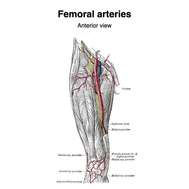
The medial circumflex femoral artery is usually a branch of the profunda femoris that arises close to its origin, usually before the origin of the lateral circumflex femoral artery. It provides blood to the femoral neck and damage to the artery or involvement of it in pathological processes may result in decreased flow and avascular necrosis of the femoral head.
On this page:
Summary
origin: profunda femoris
-
supply
adductors and hamstring muscles
branches: ascending and descending
Gross anatomy
Origin
The medial circumflex femoral artery arises from the medial aspect of the profunda femoris artery soon after it branches from the common femoral artery in the upper thigh. It travels laterally and posteriorly around the femoral neck between pectineus and the psoas tendon and supplies blood to the femoral neck along with the lateral circumflex femoral artery.
Branches
At the upper border of adductor brevis it branches into two:
ascending branch goes on to join the trochanteric anastomosis
transverse branch forms the medial limb of the cruciate anastomosis
The descending branch runs beneath adductor brevis and supplies adductor magnus before branching into three:
superficial branch
deep branch
acetabular branch
ADVERTISEMENT: Supporters see fewer/no ads
Variants
-
variant origin 1
common femoral artery: very common - 39.3%
absent: 0.6% 1
Quiz questions
References
- 1. Al-Talalwah W. The medial circumflex femoral artery origin variability and its radiological and surgical intervention significance. (2015) SpringerPlus. 4: 149. doi:10.1186/s40064-015-0881-2 - Pubmed
Incoming Links
- Pectineus muscle
- Profunda femoris branches (mnemonic)
- Gracilis muscle
- Lateral circumflex femoral artery
- Iliopsoas muscle
- Obturator externus muscle
- Trochanteric anastomosis
- Adductor minimus muscle
- Inferior gluteal artery
- Hip joint
- Superior gluteal artery
- Cruciate anastomosis
- Pubis
- Obturator artery
- Proximal femoral fractures
- Profunda femoris artery
- Femoroacetabular joint
Related articles: Anatomy: Lower limb
- skeleton of the lower limb
- joints of the lower limb
-
hip joint
- ligaments
- muscles
- additional structures
- hip joint capsule
- zona orbicularis
- iliotibial band
-
hip bursae
- anterior
- iliopsoas bursa (iliopectineal bursa)
- lateral
- subgluteal bursae
- greater trochanteric bursa (subgluteus maximus bursa)
- subgluteus medius bursa
- subgluteus minimus bursa
- gluteofemoral bursa
- subgluteal bursae
- postero-inferior
- anterior
- ossification centers
-
knee joint
- ligaments
- anterior cruciate ligament
- posterior cruciate ligament
- medial collateral ligament
- lateral collateral ligament
- meniscofemoral ligament (mnemonic)
-
posterolateral ligamentous complex
- arcuate ligament
- patellar tendon and quadriceps tendon
- anterolateral ligament
- posterior oblique ligament
- oblique popliteal ligament
- medial patellofemoral ligament
- additional structures
- extensor mechanism of the knee
- groove for the popliteus tendon
- knee bursae
- anterior bursae
- medial bursae
- lateral bursae
- posterior bursae
- knee capsule
- lateral patellar retinaculum
- medial patellar retinaculum
- menisci
- pes anserinus (mnemonic)
- ossification centers
- ligaments
- tibiofibular joints
-
ankle joint
- regional anatomy
- medial ankle
- lateral ankle
- anterior ankle
- ligaments
- medial collateral (deltoid) ligament
- lateral collateral ligament
- additional structures
- ankle bursae
- ossification centers of the ankle
- variants
- regional anatomy
- foot joints
- subtalar joint
- mid-tarsal (Chopart) joint
-
tarsometatarsal (Lisfranc) joint
- ligaments
- intermetatarsal joint
- metatarsophalangeal joint
- interphalangeal joint
- ossification centers
-
hip joint
- spaces of the lower limb
-
muscles of the lower limb
- muscles of the pelvic group
- muscles of the thigh
- muscles of the leg
- anterior compartment of the leg
- posterior compartments of the leg
- lateral compartment of the leg
- muscles of the foot
- dorsal muscles
- plantar muscles
- 1st layer
- 2nd layer
- 3rd layer
- 4th layer
- accessory muscles of the lower limb
- accessory gluteal muscles
-
accessory muscles of the ankle
- accessory peroneal muscles
- accessory flexor digitorum longus muscle
- accessory soleus muscle
- peroneocalcaneus internus muscle
- tibiocalcaneus internus muscle
- extensor hallucis capsularis tendon
- anterior fibulocalcaneus muscle
- accessory extensor digiti secundus muscle
- tibioastragalus anticus of Gruber muscle
- vascular supply of the lower limb
- arterial supply of the lower limb
- venous drainage of the lower limb
- innervation of the lower limb
- lymphatic system of the lower limb
- lymphatic pathways
- anteromedial group
- anterolateral group
- posteromedial group
- posterolateral group
- lower limb lymph nodes
- lymphatic pathways


 Unable to process the form. Check for errors and try again.