
Peroneus tertius muscle
Citation, DOI, disclosures and article data
At the time the article was created Craig Hacking had no recorded disclosures.
View Craig Hacking's current disclosuresAt the time the article was last revised Daniel J Bell had no recorded disclosures.
View Daniel J Bell's current disclosures- Fibularis tertius muscle
- Fibularis tertius
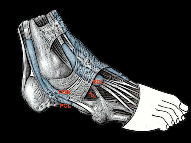
The peroneus tertius muscle, also known as fibularis tertius muscle, is a muscle of the anterior compartment of the leg, despite its name suggesting it is in the lateral compartment. It aids in dorsiflexion and eversion of the foot.
On this page:
Summary
-
origin: inferior 1/3rd of anterior surface of fibula and interosseous membrane below the extensor digitorum longus
- some authors describe the muscles as arising from the lateral margin of the extensor digitorum longus muscle
- insertion: dorsal surface of the 5th metatarsal base and with a so-called falciform extension to the superior surface of that bone
- innervation: deep peroneal nerve
- action: dorsiflexion and foot eversion
Gross anatomy
Its tendon passes anterior to the lateral malleolus (unlike peroneus brevis and longus which pass posterior), deep to the inferior extensor retinaculum to become enclosed in a common tendon sheath with the tendons of the extensor digitorum longus muscle.
ADVERTISEMENT: Supporters see fewer/no ads
Arterial supply
Variant anatomy
It may be absent or associated with other peroneus accessory muscles.
The prevalence of the peroneus tertius muscle, determined by cadaveric dissection, has been found to be 93.2% 4.
References
- 1. Chummy S. Sinnatamby. Last's Anatomy. (2018) ISBN: 9780702033940
- 2. Moore KL, Agur AMR, Dalley AF. Clinically oriented anatomy. LWW. ISBN:1451119453. Read it at Google Books - Find it at Amazon
- 3. Susan Standring. Gray's Anatomy. ISBN: 9780702052309
- 4. Yammine K, Erić M. The Fibularis (Peroneus) Tertius Muscle in Humans: A Meta-Analysis of Anatomical Studies with Clinical and Evolutionary Implications. (2017) BioMed research international. 2017: 6021707. doi:10.1155/2017/6021707 - Pubmed 4.
Incoming Links
- Anterior compartment of the leg
- Accessory peroneal muscles
- Sciatic nerve motor distribution
- Accessory muscles of the ankle
- MRI of the ankle (an approach)
- Deep peroneal nerve entrapment
- Deep peroneal nerve
- Fibula
- Anterior tarsal tunnel
- Extensor retinaculum (foot)
- Muscles of the lower limb
- Anterior lateral malleolar artery
- Metatarsal
- Anterior fibulocalcaneus muscle
Related articles: Anatomy: Lower limb
- skeleton of the lower limb
- joints of the lower limb
-
hip joint
- ligaments
- muscles
- additional structures
- hip joint capsule
- zona orbicularis
- iliotibial band
-
hip bursae
- anterior
- iliopsoas bursa (iliopectineal bursa)
- lateral
- subgluteal bursae
- greater trochanteric bursa (subgluteus maximus bursa)
- subgluteus medius bursa
- subgluteus minimus bursa
- gluteofemoral bursa
- subgluteal bursae
- postero-inferior
- anterior
- ossification centers
-
knee joint
- ligaments
- anterior cruciate ligament
- posterior cruciate ligament
- medial collateral ligament
- lateral collateral ligament
- meniscofemoral ligament (mnemonic)
-
posterolateral ligamentous complex
- arcuate ligament
- patellar tendon and quadriceps tendon
- anterolateral ligament
- posterior oblique ligament
- oblique popliteal ligament
- medial patellofemoral ligament
- additional structures
- extensor mechanism of the knee
- groove for the popliteus tendon
- knee bursae
- anterior bursae
- medial bursae
- lateral bursae
- posterior bursae
- knee capsule
- lateral patellar retinaculum
- medial patellar retinaculum
- menisci
- pes anserinus (mnemonic)
- ossification centers
- ligaments
- tibiofibular joints
-
ankle joint
- regional anatomy
- medial ankle
- lateral ankle
- anterior ankle
- ligaments
- medial collateral (deltoid) ligament
- lateral collateral ligament
- additional structures
- ankle bursae
- ossification centers of the ankle
- variants
- regional anatomy
- foot joints
- subtalar joint
- mid-tarsal (Chopart) joint
-
tarsometatarsal (Lisfranc) joint
- ligaments
- intermetatarsal joint
- metatarsophalangeal joint
- interphalangeal joint
- ossification centers
-
hip joint
- spaces of the lower limb
-
muscles of the lower limb
- muscles of the pelvic group
- muscles of the thigh
- muscles of the leg
- anterior compartment of the leg
- posterior compartments of the leg
- lateral compartment of the leg
- muscles of the foot
- dorsal muscles
- plantar muscles
- 1st layer
- 2nd layer
- 3rd layer
- 4th layer
- accessory muscles of the lower limb
- accessory gluteal muscles
-
accessory muscles of the ankle
- accessory peroneal muscles
- accessory flexor digitorum longus muscle
- accessory soleus muscle
- peroneocalcaneus internus muscle
- tibiocalcaneus internus muscle
- extensor hallucis capsularis tendon
- anterior fibulocalcaneus muscle
- accessory extensor digiti secundus muscle
- tibioastragalus anticus of Gruber muscle
- vascular supply of the lower limb
- arterial supply of the lower limb
- venous drainage of the lower limb
- innervation of the lower limb
- lymphatic system of the lower limb
- lymphatic pathways
- anteromedial group
- anterolateral group
- posteromedial group
- posterolateral group
- lower limb lymph nodes
- lymphatic pathways


 Unable to process the form. Check for errors and try again.