
Popliteal fossa
Citation, DOI, disclosures and article data
At the time the article was created Craig Hacking had no recorded disclosures.
View Craig Hacking's current disclosuresAt the time the article was last revised Mohd Ashyiraff Ilani Bin Ismail had no financial relationships to ineligible companies to disclose.
View Mohd Ashyiraff Ilani Bin Ismail's current disclosures- popliteal fossae
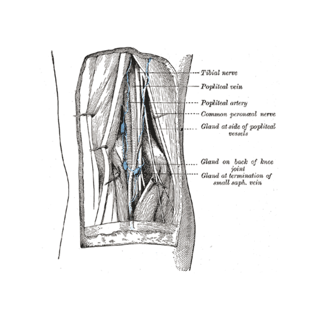
The popliteal fossa (plural: fossae) is a diamond or rhomboid-shaped fat-filled space in the posterior knee. The space is extremely dynamic, allowing for its neurovascular contents to move during the extreme range of motion produced by knee flexion and extension.
Gross anatomy
Boundaries
superolateral: medial border of biceps femoris muscle
superomedial: lateral border of semimembranosus muscle (with the tendon of semitendinosus superficial to it)
inferolateral: medial border of the lateral head of gastrocnemius, plantaris muscle
inferomedial: lateral border of the medial head of gastrocnemius
floor: (superior to inferior) popliteal surface of the femur, knee joint capsule, popliteus muscle
roof: skin, subcutaneous tissue, fascia lata
See the mnemonic here.
Contents
Arteries:
popliteal artery: deepest, gives off paired superior, middle and inferior genicular arteries 4
Veins:
popliteal vein: in between the artery and tibial nerve
short saphenous vein: ascends and pierces the roof to enter the popliteal vein in the lower half of the fossa before joining the popliteal vein
Nerves:
tibial nerve: most superficial, giving off the medial sural cutaneous nerve which descends to pierce the roof, before joining the lateral cutaneous branch to form the sural nerve4
common fibular (peroneal) nerve: runs along the lateral border, giving off the lateral cutaneous branch of the sural nerve which descends to pierce the roof in the lateral fossa gives off a communicating branch which unites with the medial branch (from the tibial nerve) to form the sural nerve4
posterior femoral cutaneous nerve: descends and pierces the roof
articular branch of obturator nerve 4
Other
fat
popliteal lymph nodes
a variable number of bursae 4
At all levels, the popliteal vein is found between the popliteal artery and the tibial nerve 2.
Related pathology
References
- 1. Moore KL, Dalley AF. Anatomy. Lippincott Williams & Wilkins. (1999) ISBN:0683061410. Read it at Google Books - Find it at Amazon
- 2. Mcminn. Last's Anatomy. Elsevier Australia. (2003) ISBN:0729537528. Read it at Google Books - Find it at Amazon
- 3. Butler P, Mitchell A, Healy JC. Applied Radiological Anatomy. Cambridge University Press. (2012) ISBN:0521766664. Read it at Google Books - Find it at Amazon
- 4. Susan Standring. Gray's Anatomy. (2020) ISBN: 9780702077050 - Google Books
Incoming Links
- Middle genicular artery
- Semimembranosus muscle
- Accessory semimembranosus muscle
- Popliteal artery
- Anterior tibial vein
- Synovial sarcoma
- Adductor canal
- Anterior tibial artery
- Sciatic neuropathy
- Posterior tibial veins
- Musculoskeletal curriculum
- Gastrocnemius muscle
- Tensor fasciae suralis muscle
- Posterior femoral cutaneous nerve
- Tibial nerve
- Linea aspera
- Baker cyst
- Crural fascia
- Popliteal vein
- Ultrasound of the knee
Related articles: Anatomy: Lower limb
- skeleton of the lower limb
- joints of the lower limb
-
hip joint
- ligaments
- muscles
- additional structures
- hip joint capsule
- zona orbicularis
- iliotibial band
-
hip bursae
- anterior
- iliopsoas bursa (iliopectineal bursa)
- lateral
- subgluteal bursae
- greater trochanteric bursa (subgluteus maximus bursa)
- subgluteus medius bursa
- subgluteus minimus bursa
- gluteofemoral bursa
- subgluteal bursae
- postero-inferior
- anterior
- ossification centers
-
knee joint
- ligaments
- anterior cruciate ligament
- posterior cruciate ligament
- medial collateral ligament
- lateral collateral ligament
- meniscofemoral ligament (mnemonic)
-
posterolateral ligamentous complex
- arcuate ligament
- patellar tendon and quadriceps tendon
- anterolateral ligament
- posterior oblique ligament
- oblique popliteal ligament
- medial patellofemoral ligament
- additional structures
- extensor mechanism of the knee
- groove for the popliteus tendon
- knee bursae
- anterior bursae
- medial bursae
- lateral bursae
- posterior bursae
- knee capsule
- lateral patellar retinaculum
- medial patellar retinaculum
- menisci
- pes anserinus (mnemonic)
- ossification centers
- ligaments
- tibiofibular joints
-
ankle joint
- regional anatomy
- medial ankle
- lateral ankle
- anterior ankle
- ligaments
- medial collateral (deltoid) ligament
- lateral collateral ligament
- additional structures
- ankle bursae
- ossification centers of the ankle
- variants
- regional anatomy
- foot joints
- subtalar joint
- mid-tarsal (Chopart) joint
-
tarsometatarsal (Lisfranc) joint
- ligaments
- intermetatarsal joint
- metatarsophalangeal joint
- interphalangeal joint
- ossification centers
-
hip joint
- spaces of the lower limb
-
muscles of the lower limb
- muscles of the pelvic group
- muscles of the thigh
- muscles of the leg
- anterior compartment of the leg
- posterior compartments of the leg
- lateral compartment of the leg
- muscles of the foot
- dorsal muscles
- plantar muscles
- 1st layer
- 2nd layer
- 3rd layer
- 4th layer
- accessory muscles of the lower limb
- accessory gluteal muscles
-
accessory muscles of the ankle
- accessory peroneal muscles
- accessory flexor digitorum longus muscle
- accessory soleus muscle
- peroneocalcaneus internus muscle
- tibiocalcaneus internus muscle
- extensor hallucis capsularis tendon
- anterior fibulocalcaneus muscle
- accessory extensor digiti secundus muscle
- tibioastragalus anticus of Gruber muscle
- vascular supply of the lower limb
- arterial supply of the lower limb
- venous drainage of the lower limb
- innervation of the lower limb
- lymphatic system of the lower limb
- lymphatic pathways
- anteromedial group
- anterolateral group
- posteromedial group
- posterolateral group
- lower limb lymph nodes
- lymphatic pathways


 Unable to process the form. Check for errors and try again.