
Quadriceps tendon
Last revised by Tariq Walizai
on 21 Aug 2024
Citation, DOI, disclosures and article data
Citation:
Gaillard F, Walizai T, Hacking C, et al. Quadriceps tendon. Reference article, Radiopaedia.org (Accessed on 20 Mar 2025) https://doi.org/10.53347/rID-1944
rID:
1944
Article created:
Disclosures:
At the time the article was created Frank Gaillard had no recorded disclosures.
View Frank Gaillard's current disclosures
Last revised:
21 Aug 2024,
Tariq Walizai
Disclosures:
At the time the article was last revised Tariq Walizai had no financial relationships to ineligible companies to disclose.
View Tariq Walizai's current disclosures
Revisions:
11 times, by
9 contributors -
see full revision history and disclosures
Systems:
Sections:
Synonyms:
- Quadriceps tendons
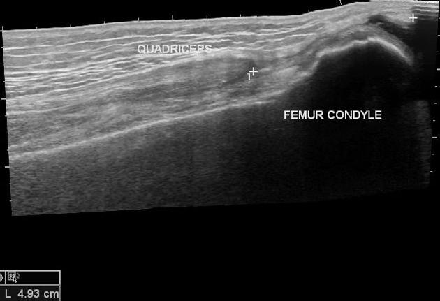
The quadriceps tendon is a thick tendon extending to the patella made up of contributions from all four quadriceps muscles. It classically has a trilaminar structure:
superficial layer: rectus femoris
middle layer: vastus medialis, vastus lateralis
deep layer: vastus intermedius
It continues distal to the patella as the patellar tendon.
See also
References
- 1. Zeiss J, Saddemi SR, Ebraheim NA. MR imaging of the quadriceps tendon: normal layered configuration and its importance in cases of tendon rupture. AJR Am J Roentgenol. 1992;159 (5): 1031-4. AJR Am J Roentgenol (abstract) - Pubmed citation
Incoming Links
Articles:
- Joint effusion
- Medial patellar retinaculum
- Superior lateral genicular artery
- Vastus intermedius muscle
- Sinding-Larsen-Johansson disease
- Vastus lateralis muscle
- Quadriceps muscles
- Quadriceps tendon rupture
- Patella
- Quadriceps injury
- Anterior suprapatellar fat pad impingement syndrome
- Patellar tendon
- Vastus medialis muscle
- Lateral patellar retinaculum
- Extensor mechanism of the knee
- Descending genicular artery
- Anterior suprapatellar fat pad
- Quadriceps tendinopathy
- Rectus femoris muscle
- Mucoid change
Cases:
- Quadriceps tendon partial tear
- Quadriceps tendon tear
- Quadriceps tendon rupture
- Lipohaemarthrosis and hemarthrosis (illustrations)
- Quadriceps fat pad impingement syndrome
- Deep infrapatellar bursitis
- Lateral collateral ligament calcification - knee
- Knee ligaments (Gray's illustration)
- Patellar fracture
- Chondromalacia patellae
- Soft tissue haemangioma - knee
- Quadriceps tendon partial tear
- Suture anchors - quadriceps tendon reimplantation
- Quadriceps tendon insertion rupture
- Discoid medial meniscus with horizontal tear
- Rectus femoris injury - distal myotendinous junction rupture
- Quadriceps muscle partial rupture and lipohaemarthrosis
- MRI knee - sagittal (anatomy quiz)
- Patellar enthesopathy
- Normal radiographic anatomy of the knee
Multiple choice questions:
Related articles: Anatomy: Lower limb
- skeleton of the lower limb
- joints of the lower limb
-
hip joint
- ligaments
- muscles
- additional structures
- hip joint capsule
- zona orbicularis
- iliotibial band
-
hip bursae
- anterior
- iliopsoas bursa (iliopectineal bursa)
- lateral
- subgluteal bursae
- greater trochanteric bursa (subgluteus maximus bursa)
- subgluteus medius bursa
- subgluteus minimus bursa
- gluteofemoral bursa
- subgluteal bursae
- postero-inferior
- anterior
- ossification centers
-
knee joint
- ligaments
- anterior cruciate ligament
- posterior cruciate ligament
- medial collateral ligament
- lateral collateral ligament
- meniscofemoral ligament (mnemonic)
-
posterolateral ligamentous complex
- arcuate ligament
- patellar tendon and quadriceps tendon
- anterolateral ligament
- posterior oblique ligament
- oblique popliteal ligament
- medial patellofemoral ligament
- additional structures
- extensor mechanism of the knee
- groove for the popliteus tendon
- knee bursae
- anterior bursae
- medial bursae
- lateral bursae
- posterior bursae
- knee capsule
- lateral patellar retinaculum
- medial patellar retinaculum
- menisci
- pes anserinus (mnemonic)
- ossification centers
- ligaments
- tibiofibular joints
-
ankle joint
- regional anatomy
- medial ankle
- lateral ankle
- anterior ankle
- ligaments
- medial collateral (deltoid) ligament
- lateral collateral ligament
- additional structures
- ankle bursae
- ossification centers of the ankle
- variants
- regional anatomy
- foot joints
- subtalar joint
- mid-tarsal (Chopart) joint
-
tarsometatarsal (Lisfranc) joint
- ligaments
- intermetatarsal joint
- metatarsophalangeal joint
- interphalangeal joint
- ossification centers
-
hip joint
- spaces of the lower limb
-
muscles of the lower limb
- muscles of the pelvic group
- muscles of the thigh
- muscles of the leg
- anterior compartment of the leg
- posterior compartments of the leg
- lateral compartment of the leg
- muscles of the foot
- dorsal muscles
- plantar muscles
- 1st layer
- 2nd layer
- 3rd layer
- 4th layer
- accessory muscles of the lower limb
- accessory gluteal muscles
-
accessory muscles of the ankle
- accessory peroneal muscles
- accessory flexor digitorum longus muscle
- accessory soleus muscle
- peroneocalcaneus internus muscle
- tibiocalcaneus internus muscle
- extensor hallucis capsularis tendon
- anterior fibulocalcaneus muscle
- accessory extensor digiti secundus muscle
- tibioastragalus anticus of Gruber muscle
- vascular supply of the lower limb
- arterial supply of the lower limb
- venous drainage of the lower limb
- innervation of the lower limb
- lymphatic system of the lower limb
- lymphatic pathways
- anteromedial group
- anterolateral group
- posteromedial group
- posterolateral group
- lower limb lymph nodes
- lymphatic pathways


 Unable to process the form. Check for errors and try again.