
Rectus femoris muscle
Citation, DOI, disclosures and article data
At the time the article was created a b had no recorded disclosures.
View a b's current disclosuresAt the time the article was last revised Roy Martin Spires had no financial relationships to ineligible companies to disclose.
View Roy Martin Spires's current disclosures- Rectus femoris muscles
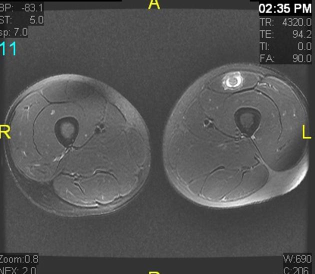
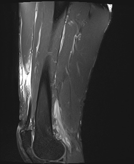
The rectus femoris muscle is one of four quadriceps muscles in the anterior compartment of the thigh. It is distinct from the other quadriceps muscles (vastus medialis, vastus intermedius, vastus lateralis) in that it crosses both the hip and knee joints 1.
On this page:
Summary
-
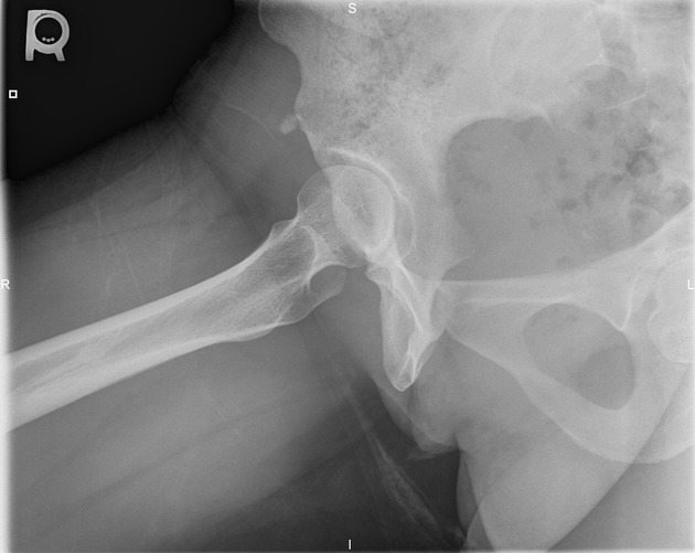
origin(s): consists of two proximal heads which form a conjoined tendon 1,5
direct/straight head: anterior inferior iliac spine (AIIS) of the ilium
indirect/reflected head: superior acetabular ridge
insertion: quadriceps tendon
action: flexes the thigh at the hip joint and extends the leg at the knee joint
arterial supply: descending branch of the lateral circumflex femoral artery
innervation: femoral nerve
Gross anatomy
The rectus femoris has two heads with separate origins 5,6:
direct/straight head: AIIS
indirect/reflected head: superior acetabular ridge
These two tendons merge ~1 cm below their origin to form the conjoined tendon with two components 5,6:
superficial/anterior component: blends more so with the anterior fascia
deep/posterior component: forms the central tendon with a long myotendinous junction
There is an intermingling of ~15% of muscle fibers related to each origin, and rectus femoris can be thought of as two muscles 7. The distal myotendinous junction forms a short free tendon that joins with the vastus tendons to form the quadriceps tendon.
Radiographic features
Ultrasound
Patient in a supine position in hip extension and probe placed of the AIIS in a longitudinal plane 4:
direct head: seen directly at the insertion on AIIS
indirect head: hypoechoic appearance due to oblique course
Related pathology
Rectus femoris muscle injuries in athletes include 4-6:
direct and indirect head or free tendon injury
proximal myotendinous junction injury
distal myotendinous junction injury
Other pathology includes:
Quiz questions
References
- 1. Gray's Anatomy for Students: With STUDENT CONSULT Online Access, 3e. Churchill Livingstone. ISBN:0702051314. Read it at Google Books - Find it at Amazon
- 2. Clinically Oriented Anatomy. Lippincott Williams & Wilkins. ISBN:1451119453. Read it at Google Books - Find it at Amazon
- 3. Last's Anatomy. Churchill Livingstone. ISBN:0702033944. Read it at Google Books - Find it at Amazon
- 4. Lungu E, Michaud J, Bureau NJ. US Assessment of Sports-related Hip Injuries. (2018) Radiographics : a review publication of the Radiological Society of North America, Inc. 38 (3): 867-889. doi:10.1148/rg.2018170104 - Pubmed
- 5. Soterios Gyftopoulos, Zehava Sadka Rosenberg, Mark E. Schweitzer, Marcelo Bordalo-Rodrigues. Normal Anatomy and Strains of the Deep Musculotendinous Junction of the Proximal Rectus Femoris: MRI Features. (2012) American Journal of Roentgenology. 190 (3): W182-6. doi:10.2214/AJR.07.2947 - Pubmed
- 6. Brukner P, Connell D. 'Serious thigh muscle strains': beware the intramuscular tendon which plays an important role in difficult hamstring and quadriceps muscle strains. (2016) British journal of sports medicine. 50 (4): 205-8. doi:10.1136/bjsports-2015-095136 - Pubmed
- 7. Hasselman CT, Best TM, Hughes C, Martinez S, Garrett WE. An explanation for various rectus femoris strain injuries using previously undescribed muscle architecture. (1995) The American journal of sports medicine. 23 (4): 493-9. doi:10.1177/036354659502300421 - Pubmed
Incoming Links
- Rectus femoris heterotopic ossification
- Anterior inferior iliac spine
- Duchenne muscular dystrophy
- Vastus intermedius muscle
- Anterior inferior iliac spine avulsion injury
- Quadriceps muscles
- Ilium
- Quadriceps injury
- Patellar tendon
- Apophyseal avulsion fractures of the pelvis and hip
- Muscle tear
- Vastus medialis muscle
- Anterior compartment of the thigh
- Descending genicular artery
- Muscles of the lower limb
- Inclusion body myositis
- Quadriceps tendon
- Prepatellar quadriceps continuation
- Rectus femoris muscle injury
- Intramuscular degloving injury
- Myositis ossificans of rectus femoris
- Anterior inferior iliac spine avulsion injury
- Complete proximal rectus femoris tendon tear
- Bilateral rectus femoris tendon ossification - indirect head
- Rectus femoris muscle lipoma
- Proximal rectus femoris tendon tear
- Rectus femoris heterotopic ossification
- Psoas abscess
- Subspine impingement of the hip
- Rectus femoris origin tendon ossification
- Undifferentiated pleomorphic sarcoma
- Right rectus femoris strain
- Rectus femoris muscle injury
- Rectus femoris avulsion injury
- Rectus femoris muscle injury
- Rectus femoris muscle injury
- Proximal rectus femoris tendon tear
- Rectus femoris injury - intramuscular degloving
- Intramuscular lipoma - rectus femoris muscle
- Rectus femoris injury - distal myotendinous junction rupture
Related articles: Anatomy: Lower limb
- skeleton of the lower limb
- joints of the lower limb
-
hip joint
- ligaments
- muscles
- additional structures
- hip joint capsule
- zona orbicularis
- iliotibial band
-
hip bursae
- anterior
- iliopsoas bursa (iliopectineal bursa)
- lateral
- subgluteal bursae
- greater trochanteric bursa (subgluteus maximus bursa)
- subgluteus medius bursa
- subgluteus minimus bursa
- gluteofemoral bursa
- subgluteal bursae
- postero-inferior
- anterior
- ossification centers
-
knee joint
- ligaments
- anterior cruciate ligament
- posterior cruciate ligament
- medial collateral ligament
- lateral collateral ligament
- meniscofemoral ligament (mnemonic)
-
posterolateral ligamentous complex
- arcuate ligament
- patellar tendon and quadriceps tendon
- anterolateral ligament
- posterior oblique ligament
- oblique popliteal ligament
- medial patellofemoral ligament
- additional structures
- extensor mechanism of the knee
- groove for the popliteus tendon
- knee bursae
- anterior bursae
- medial bursae
- lateral bursae
- posterior bursae
- knee capsule
- lateral patellar retinaculum
- medial patellar retinaculum
- menisci
- pes anserinus (mnemonic)
- ossification centers
- ligaments
- tibiofibular joints
-
ankle joint
- regional anatomy
- medial ankle
- lateral ankle
- anterior ankle
- ligaments
- medial collateral (deltoid) ligament
- lateral collateral ligament
- additional structures
- ankle bursae
- ossification centers of the ankle
- variants
- regional anatomy
- foot joints
- subtalar joint
- mid-tarsal (Chopart) joint
-
tarsometatarsal (Lisfranc) joint
- ligaments
- intermetatarsal joint
- metatarsophalangeal joint
- interphalangeal joint
- ossification centers
-
hip joint
- spaces of the lower limb
-
muscles of the lower limb
- muscles of the pelvic group
- muscles of the thigh
- muscles of the leg
- anterior compartment of the leg
- posterior compartments of the leg
- lateral compartment of the leg
- muscles of the foot
- dorsal muscles
- plantar muscles
- 1st layer
- 2nd layer
- 3rd layer
- 4th layer
- accessory muscles of the lower limb
- accessory gluteal muscles
-
accessory muscles of the ankle
- accessory peroneal muscles
- accessory flexor digitorum longus muscle
- accessory soleus muscle
- peroneocalcaneus internus muscle
- tibiocalcaneus internus muscle
- extensor hallucis capsularis tendon
- anterior fibulocalcaneus muscle
- accessory extensor digiti secundus muscle
- tibioastragalus anticus of Gruber muscle
- vascular supply of the lower limb
- arterial supply of the lower limb
- venous drainage of the lower limb
- innervation of the lower limb
- lymphatic system of the lower limb
- lymphatic pathways
- anteromedial group
- anterolateral group
- posteromedial group
- posterolateral group
- lower limb lymph nodes
- lymphatic pathways


 Unable to process the form. Check for errors and try again.