
Suprapatellar bursa
Citation, DOI, disclosures and article data
Citation:
Gilcrease-Garcia B, Hacking C, Bell D, et al. Suprapatellar bursa. Reference article, Radiopaedia.org (Accessed on 21 Mar 2025) https://doi.org/10.53347/rID-62254
rID:
62254
Article created:
8 Aug 2018,
Brian Gilcrease-Garcia ◉
Disclosures:
At the time the article was created Brian Gilcrease-Garcia had no recorded disclosures.
View Brian Gilcrease-Garcia's current disclosures
Last revised:
Disclosures:
At the time the article was last revised Craig Hacking had no recorded disclosures.
View Craig Hacking's current disclosures
Revisions:
8 times, by
7 contributors -
see full revision history and disclosures
Systems:
Sections:
Tags:
Synonyms:
- Suprapatellar pouch
- Suprapatellar bursae
- Supra-patellar bursa
- Suprapatellar recess
- Suprapatellar pouches
- Suprapatellar recesses
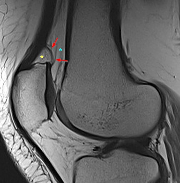
The suprapatellar bursa, also known as the suprapatellar recess or suprapatellar pouch, is one of several bursae of the knee. It is located proximal to the knee joint, between the prefemoral and suprapatellar fat pads. As with all bursae, its purpose is to reduce friction between moving structures.
In most (~85%) people, the suprapatellar bursa communicates with the knee joint proper. Thus, it is useful to assess for bursal distension on x-ray, as this generally indicates the presence of a knee effusion.
Variant anatomy
- the suprapatellar bursa does not communicate with the knee joint in ~15% of people, remaining separated by an embryonic septum 2,3
Related pathology
References
- 1. Gray, Henry. Anatomy of the Human Body: 7b. The Knee Joint. Philadelphia: Lea & Febiger, 1918; Bartleby.com, 2000. Available at Bartleby.com
- 2. Steinbach LS, Stevens KJ. Imaging of cysts and bursae about the knee. (2013) Radiologic clinics of North America. 51 (3): 433-54. doi:10.1016/j.rcl.2012.10.005 - Pubmed
- 3. Zidorn T, Schäfer H. Morphologic variants of the proximal knee-joint cavity. An anatomical and radiological study. (1992) Surgical and radiologic anatomy : SRA. 14 (2): 141-6. Pubmed
Incoming Links
Articles:
Cases:
- Gout
- Prepatellar bursitis
- Osteomyelitis - tibia
- Tibial plateau ORIF - Schatzker type III fracture
- Tibial eminence avulsion fracture - Meyers and McKeever type 3b
- Superficial infrapatellar bursitis
- Quadriceps fat pad impingement syndrome
- Synovial osteochondromatosis
- Deep infrapatellar bursitis
- Lateral collateral ligament calcification - knee
- Gossypiboma - knee
- Knee bursae (Gray's illustration)
- Chronic osteochondral fracture - lateral femoral trochlea
- Suprapatellar bursa - normal (MRI)
- Tibial plateau fracture: Schatzker type V
- Prepatellar bursitis
- Lipoma arborescens
- Lipoma arborescens
- Synovial osteochondromatosis
- Lipoma arborescens
Multiple choice questions:
Related articles: Anatomy: Lower limb
- skeleton of the lower limb
- joints of the lower limb
-
hip joint
- ligaments
- muscles
- additional structures
- hip joint capsule
- zona orbicularis
- iliotibial band
-
hip bursae
- anterior
- iliopsoas bursa (iliopectineal bursa)
- lateral
- subgluteal bursae
- greater trochanteric bursa (subgluteus maximus bursa)
- subgluteus medius bursa
- subgluteus minimus bursa
- gluteofemoral bursa
- subgluteal bursae
- postero-inferior
- anterior
- ossification centers
-
knee joint
- ligaments
- anterior cruciate ligament
- posterior cruciate ligament
- medial collateral ligament
- lateral collateral ligament
- meniscofemoral ligament (mnemonic)
-
posterolateral ligamentous complex
- arcuate ligament
- patellar tendon and quadriceps tendon
- anterolateral ligament
- posterior oblique ligament
- oblique popliteal ligament
- medial patellofemoral ligament
- additional structures
- extensor mechanism of the knee
- groove for the popliteus tendon
- knee bursae
- anterior bursae
- medial bursae
- lateral bursae
- posterior bursae
- knee capsule
- lateral patellar retinaculum
- medial patellar retinaculum
- menisci
- pes anserinus (mnemonic)
- ossification centers
- ligaments
- tibiofibular joints
-
ankle joint
- regional anatomy
- medial ankle
- lateral ankle
- anterior ankle
- ligaments
- medial collateral (deltoid) ligament
- lateral collateral ligament
- additional structures
- ankle bursae
- ossification centers of the ankle
- variants
- regional anatomy
- foot joints
- subtalar joint
- mid-tarsal (Chopart) joint
-
tarsometatarsal (Lisfranc) joint
- ligaments
- intermetatarsal joint
- metatarsophalangeal joint
- interphalangeal joint
- ossification centers
-
hip joint
- spaces of the lower limb
-
muscles of the lower limb
- muscles of the pelvic group
- muscles of the thigh
- muscles of the leg
- anterior compartment of the leg
- posterior compartments of the leg
- lateral compartment of the leg
- muscles of the foot
- dorsal muscles
- plantar muscles
- 1st layer
- 2nd layer
- 3rd layer
- 4th layer
- accessory muscles of the lower limb
- accessory gluteal muscles
-
accessory muscles of the ankle
- accessory peroneal muscles
- accessory flexor digitorum longus muscle
- accessory soleus muscle
- peroneocalcaneus internus muscle
- tibiocalcaneus internus muscle
- extensor hallucis capsularis tendon
- anterior fibulocalcaneus muscle
- accessory extensor digiti secundus muscle
- tibioastragalus anticus of Gruber muscle
- vascular supply of the lower limb
- arterial supply of the lower limb
- venous drainage of the lower limb
- innervation of the lower limb
- lymphatic system of the lower limb
- lymphatic pathways
- anteromedial group
- anterolateral group
- posteromedial group
- posterolateral group
- lower limb lymph nodes
- lymphatic pathways


 Unable to process the form. Check for errors and try again.