
Sural nerve
Citation, DOI, disclosures and article data
At the time the article was created Henry Smith had no recorded disclosures.
View Henry Smith's current disclosuresAt the time the article was last revised Arlene Campos had no financial relationships to ineligible companies to disclose.
View Arlene Campos's current disclosuresThe sural nerve ( in Latin Sura means Calf) is a sensory nerve of the lower limb formed by the union of sural branch of the tibial nerve and the communicating sural branch of the common fibular nerve supplying sensation to the lower lateral aspect of the calf and foot.
On this page:
Gross anatomy
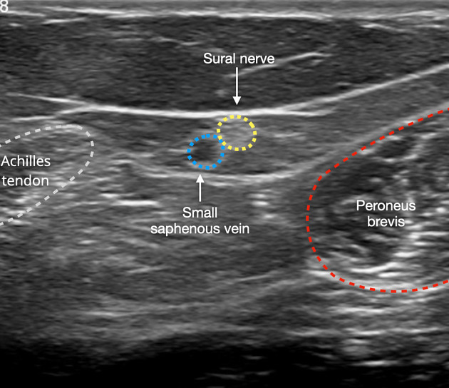
It travels within subcutaneous tissue adjacent to the small saphenous vein in the lower posterolateral calf. The nerve descends and curls forward behind the lateral malleolus and becomes the lateral dorsal cutaneous nerve. Usually comprising the nerve roots S1 and S2 it provides sensation to the lower lateral calf, lateral ankle, lateral foot and some of the 5th digit 1-3.
Variant anatomy
The nerve has a variable anatomical formation. The most common of which is via the union of the medial sural cutaneous nerve (a branch of the proximal tibial nerve) and the peroneal communicating nerve from the lateral sural cutaneous nerve (a branch of the common peroneal nerve). The nerve may be formed solely from either of these branches 2,3.
ADVERTISEMENT: Supporters see fewer/no ads
Related pathology
Iatrogenic injury to the sural nerve can occur with operative procedures involving the ankle, Achilles tendon and the small saphenous vein. Injury to the nerve is well tolerated, and for this reason, the is often used for nerve grafting or biopsy 4,5.
Quiz questions

References
- 1. Staniforth P & Fisher T. The Effects of Sural Nerve Excision in Autogenous Nerve Grafting. Hand. 1978;10(2):187-90. doi:10.1016/s0072-968x(78)80012-6 - Pubmed
- 2. Juan M Bilbao, Robert E Schmidt. Biopsy Diagnosis of Peripheral Neuropathy. (2014) ISBN: 9783319073101 - Google Books
- 3. Aktan Ikiz Z, Uçerler H, Bilge O. The Anatomic Features of the Sural Nerve with an Emphasis on Its Clinical Importance. Foot Ankle Int. 2005;26(7):560-7. doi:10.1177/107110070502600712 - Pubmed
- 4. Riedl O & Frey M. Anatomy of the Sural Nerve: Cadaver Study and Literature Review. Plast Reconstr Surg. 2013;131(4):802-10. doi:10.1097/PRS.0b013e3182818cd4 - Pubmed
- 5. Susan Standring. Gray's Anatomy. (2008) ISBN: 9780443066849 - Google Books
Incoming Links
Related articles: Anatomy: Lower limb
- skeleton of the lower limb
- joints of the lower limb
-
hip joint
- ligaments
- muscles
- additional structures
- hip joint capsule
- zona orbicularis
- iliotibial band
-
hip bursae
- anterior
- iliopsoas bursa (iliopectineal bursa)
- lateral
- subgluteal bursae
- greater trochanteric bursa (subgluteus maximus bursa)
- subgluteus medius bursa
- subgluteus minimus bursa
- gluteofemoral bursa
- subgluteal bursae
- postero-inferior
- anterior
- ossification centers
-
knee joint
- ligaments
- anterior cruciate ligament
- posterior cruciate ligament
- medial collateral ligament
- lateral collateral ligament
- meniscofemoral ligament (mnemonic)
-
posterolateral ligamentous complex
- arcuate ligament
- patellar tendon and quadriceps tendon
- anterolateral ligament
- posterior oblique ligament
- oblique popliteal ligament
- medial patellofemoral ligament
- additional structures
- extensor mechanism of the knee
- groove for the popliteus tendon
- knee bursae
- anterior bursae
- medial bursae
- lateral bursae
- posterior bursae
- knee capsule
- lateral patellar retinaculum
- medial patellar retinaculum
- menisci
- pes anserinus (mnemonic)
- ossification centers
- ligaments
- tibiofibular joints
-
ankle joint
- regional anatomy
- medial ankle
- lateral ankle
- anterior ankle
- ligaments
- medial collateral (deltoid) ligament
- lateral collateral ligament
- additional structures
- ankle bursae
- ossification centers of the ankle
- variants
- regional anatomy
- foot joints
- subtalar joint
- mid-tarsal (Chopart) joint
-
tarsometatarsal (Lisfranc) joint
- ligaments
- intermetatarsal joint
- metatarsophalangeal joint
- interphalangeal joint
- ossification centers
-
hip joint
- spaces of the lower limb
-
muscles of the lower limb
- muscles of the pelvic group
- muscles of the thigh
- muscles of the leg
- anterior compartment of the leg
- posterior compartments of the leg
- lateral compartment of the leg
- muscles of the foot
- dorsal muscles
- plantar muscles
- 1st layer
- 2nd layer
- 3rd layer
- 4th layer
- accessory muscles of the lower limb
- accessory gluteal muscles
-
accessory muscles of the ankle
- accessory peroneal muscles
- accessory flexor digitorum longus muscle
- accessory soleus muscle
- peroneocalcaneus internus muscle
- tibiocalcaneus internus muscle
- extensor hallucis capsularis tendon
- anterior fibulocalcaneus muscle
- accessory extensor digiti secundus muscle
- tibioastragalus anticus of Gruber muscle
- vascular supply of the lower limb
- arterial supply of the lower limb
- venous drainage of the lower limb
- innervation of the lower limb
- lymphatic system of the lower limb
- lymphatic pathways
- anteromedial group
- anterolateral group
- posteromedial group
- posterolateral group
- lower limb lymph nodes
- lymphatic pathways


 Unable to process the form. Check for errors and try again.