
The tibia (plural: tibiae) is the largest bone of the leg and contributes to the knee and ankle joints. (shin- or shank-bone are lay terms). It is medial to and much stronger than the fibula, exceeded in length only by the femur.
On this page:
Gross anatomy
Osteology
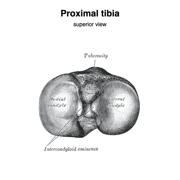
The superior tibial surface, known as the tibial plateau 2, consists of medial and lateral articular facets that articulate with the respective medial and lateral femoral condyles, forming the tibiofemoral joint of the knee 1.
The intercondylar area separates the medial and lateral articular surfaces with a central intercondylar eminence from which the medial and lateral tubercles arise 1. The intercondylar area is divided into the anterior and posterior areas. The anterior intercondylar area houses the attachment of the anterior cruciate ligament, the anterior horn of the medial meniscus, and a small part of the anterior horn of the lateral meniscus. The posterior intercondylar area slopes downwards and posteriorly, exhibiting a depression posterior to the medial intercondylar tubercle for the posterior horn of the lateral meniscus attachment and a smooth tapering ridge for the posterior cruciate ligament attachment 1.
The tibial tuberosity is a bony projection of the area where the anterior condylar surface merges. It receives the patellar tendon attachment and is separated from the skin by the subcutaneous infrapatellar bursa. Gerdy's tubercle is located at the anterolateral aspect of the proximal tibia, where the iliotibial band attaches.
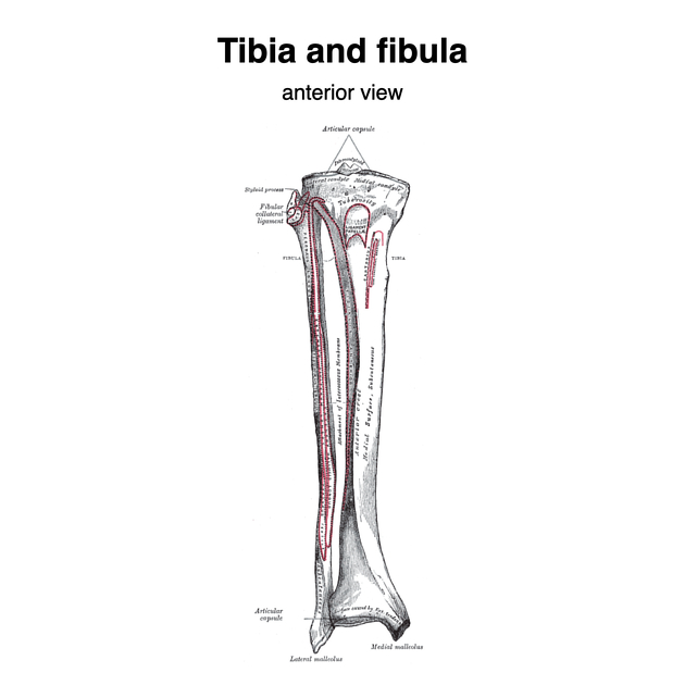
The tibial diaphysis is triangular in cross-section and has medial, lateral, and posterior surfaces separated by the anterior, lateral (interosseous), and medial borders. The shaft is thinnest at the junction of the middle and distal thirds.
The slightly expanded end of the tibia is rotated laterally (tibial torsion) and has five surfaces; anterior, posterior, medial, lateral and distal. The lateral surface exhibits a triangular notch which attaches to the fibula.
Articulations
-
proximal
medial and lateral facets of tibial condyles with the medial and lateral femoral condyles (forming the tibiofemoral joint) 1
fibular articular facet of the lateral tibia condyle with the fibular head (forming the proximal tibiofibular joint) 1
-
distal
sellar surface of tibia with the dome of talus (talocrural joint/tibiotalar joint)
distal tibia with the lateral malleolus of the distal fibula (distal tibiofibular joint/syndesmosis)
Attachments
Musculotendinous
-
anterior
tensor fascia latae: Gerdy's tubercle via the iliotibial band
quadriceps femoris: tibial tuberosity via the patellar tendon
sartorius, gracilis, semitendinosus form the pes anserinus with an underlying pes anserine bursa: anteriomedial proximal tibia
-
medial
semimembranosus (horizontal head): medial condyle
-
lateral
tibialis anterior: lateral tibia
extensor digitorum longus: lateral tibial shaft
-
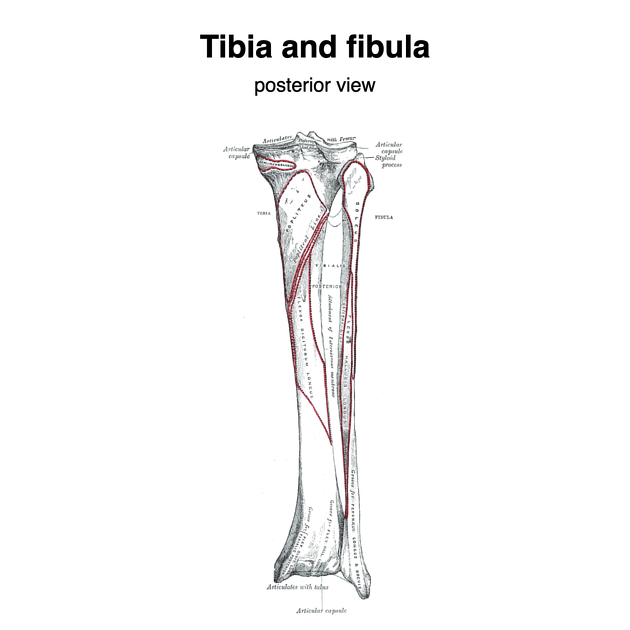
posterior
popliteus: posterior tibia above the soleal line
soleus: posterior tibia on the soleal line
flexor digitorum longus (medial): posterior tibia distal to soleal line
tibialis posterior muscle (lateral): posterior tibia distal to soleal line
Ligamentous
-
proximal
patellar tendon
anterior and posterior proximal tibiofibular ligaments
-
distal
anterior and posterior tibiofibular ligaments
anterior and posterior tibiofibular ligaments
Relations
At the medial malleolus, three tendons pass posteriorly (in order of anterior to posterior): tibialis posterior, flexor digitorum longus and flexor hallucis longus.
Arterial supply
The nutrient artery (a branch of the posterior tibial artery) enters through the nutrient foramen at the level of soleal line and is the major arterial supply for the tibia. The proximal metaphysis receives supply from the genicular arterial anastomosis, and the periosteum via the anterior tibial artery as it branches to form the periosteal arteries. Arterial anastomosis at ankle supplies the distal end of tibia.
Nerve supply
The tibia is proximally innervated by branches supplying the knee joint, and distally by those supplying the ankle. The periosteum of the diaphysis receives nerve twigs from the overlying muscles attaching to the tibia.
Variant anatomy
Development
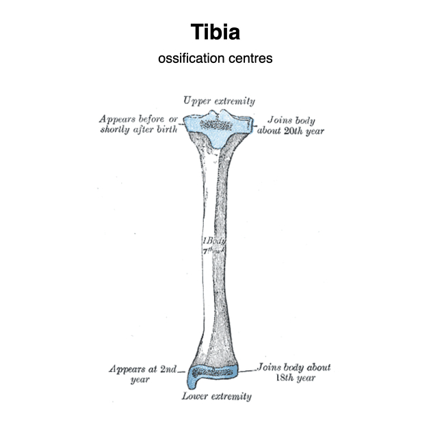
Ossification
The tibia ossifies from three centers; one in the diaphysis and one at each of the proximal and distal epiphysis.
The diaphyseal ossification center appears at the seventh week antenatally. The proximal ossification center appears at birth and fuses in the sixteenth year in females and the eighteenth year in males. The distal ossification center appears at the first year of life and joins the shaft at the fifteenth year in females, and the seventeenth year in males.
The medial malleolus is merely an extension from the distal epiphysis and ossifies at seventh year of life.


 Unable to process the form. Check for errors and try again.