
The transverse colon is the longest and most mobile part of the large intestine. It measures up to 45 cm in length.
On this page:
Gross anatomy
The transverse colon is the continuation of the ascending colon from the right colic flexure. It passes from the right to left hypochondrium in a downward convex path crossing both the epigastric and umbilical zones. In the left hypochondrium, it curves sharply on itself beneath the lower end of the spleen, forming the left colic flexure where it continues as the descending colon.
It is almost completely invested by peritoneum, and is connected to the inferior border of the pancreas by a large and wide duplicature of that membrane, the transverse mesocolon. The gastrocolic ligament also attaches the transverse colon to the stomach. The phrenicocolic ligament attaches to the left colic flexure and connects it to the diaphragm, with variable connection to the inferior aspect of the spleen.
Relations
superiorly (right to left): liver, gallbladder, greater curvature of the stomach, spleen
inferiorly: small bowel
anteriorly: greater omentum and anterior abdominal wall
posteriorly (right to left): 2nd part duodenum, head and body of pancreas, small bowel
Arterial supply
middle colic artery (branch of superior mesenteric artery) supplies proximal two-thirds
ascending branch of left colic artery (branch of inferior mesenteric artery) supplies distal one-third
Venous drainage
via similarly named veins to splenic vein to the portal venous system
Innervation
sympathetic: superior mesenteric plexus and inferior mesenteric plexus
parasympathetic: derived from pelvic splanchnic nerves (S2-S4)
Lymphatic drainage
Lymphatics accompany vessels and drain to paracolic nodes, and to the superior mesenteric group (proximal two-thirds) and inferior mesenteric group (distal two-thirds).
Radiographic features
Fluoroscopy
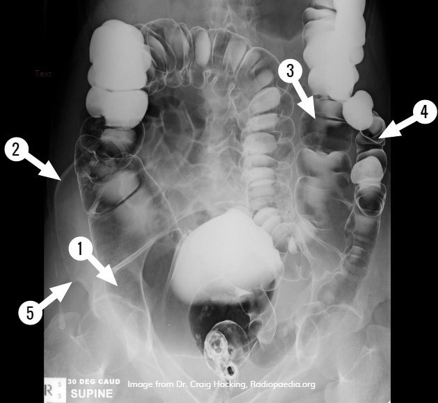
Double contrast barium enemas provided good anatomical detail from the rectum to the cecum. The patient may need to be rolled into various positions to get the barium sulfate contrast medium to coat the lumen of the colon.


 Unable to process the form. Check for errors and try again.