
Anterior angulation of the coccyx
Citation, DOI, disclosures and article data
At the time the article was created Usman Bashir had no recorded disclosures.
View Usman Bashir's current disclosuresAt the time the article was last revised Ashesh Ishwarlal Ranchod had no financial relationships to ineligible companies to disclose.
View Ashesh Ishwarlal Ranchod's current disclosures- Forward angulation of the coccyx
- Anterior deformity of the coccyx
- Anterior angulation of coccyx
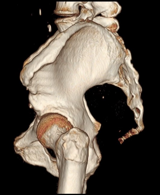
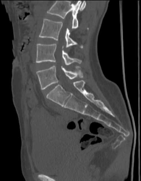
Anterior angulation of the coccyx may be a normal variant but poses a diagnostic challenge for those considering coccygeal trauma 1.
On this page:
Classification
Six types of coccyx have been described initially by Postacchini and Massobrio and later modified by Nathan et al. 2,3:
type I: the coccyx is curved slightly forward, with its apex pointing caudally (~70%)
type II: the coccyx is curved more markedly anteriorly, with its apex pointing straight forward (~15%)
type III: the coccyx is sharply angulated forward between the first and second or the second and third coccygeal segments (~5%), or less commonly due to angulation at the S5 segment
type IV: the coccyx is subluxed anteriorly at the level of the sacrococcygeal joint or at the level of the first or second intercoccygeal joints (~10%)
type V: the coccyx is retroverted or has a posterior spicule
type VI: the coccyx is scoliotic (lateral deviation)
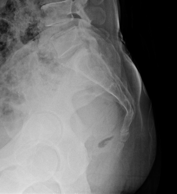
Radiographic features
The intercoccygeal angle is a useful radiological assessment to evaluate the anterior angulation of the coccyx and its deformity.
Related pathology
Patients with a type II-IV coccyx are more prone to develop idiopathic coccygodynia than those with a type I configuration ref. Partial or total coccygectomy usually provides relief in these cases ref.
Quiz questions
References
- 1. Postacchini F, Massobrio M. Idiopathic coccygodynia. Analysis of fifty-one operative cases and a radiographic study of the normal coccyx. J Bone Joint Surg Am. 1983;65 (8): 1116-24. J Bone Joint Surg Am (link) - Pubmed citation
- 2. Skalski M, Matcuk G, Patel D, Tomasian A, White E, Gross J. Imaging Coccygeal Trauma and Coccydynia. Radiographics. 2020;40(4):1090-106. doi:10.1148/rg.2020190132 - Pubmed
- 3. Nathan S, Fisher B, Roberts C. Coccydynia. The Journal of Bone and Joint Surgery British Volume. 2010;92-B(12):1622-7. doi:10.1302/0301-620x.92b12.25486 - Pubmed
Incoming Links
- Coccydynia
- Intercoccygeal angle (illustration)
- Anterior angulation of the coccyx and bilateral accessory sacroiliac joints
- Anterior angulation of the coccyx
- Anterior angulation of coccyx - type I
- Anterior angulation of the coccyx - type IV
- Anteriorly angulated coccyx
- Anterior angulation of the coccyx
- Intercoccygeal angle measurement
- Forward angulation type III coccyx
- Coccydynia
- Anterior angulation of the coccyx
Related articles: Anatomy: Abdominopelvic
- skeleton of the abdomen and pelvis
- muscles of the abdomen and pelvis
- spaces of the abdomen and pelvis
- anterior abdominal wall
- posterior abdominal wall
- abdominal cavity
- pelvic cavity
- perineum
- abdominal and pelvic viscera
- gastrointestinal tract
- spleen
- hepatobiliary system
-
endocrine system
-
adrenal gland
- adrenal vessels
- chromaffin cells
- variants
- pancreas
- organs of Zuckerkandl
-
adrenal gland
-
urinary system
-
kidney
- renal pelvis
- renal sinus
- avascular plane of Brodel
-
variants
- number
- fusion
- location
- shape
- ureter
- urinary bladder
- urethra
- embryology
-
kidney
- male reproductive system
-
female reproductive system
- vulva
- vagina
- uterus
- adnexa
- Fallopian tubes
- ovaries
- broad ligament (mnemonic)
- variant anatomy
- embryology
- blood supply of the abdomen and pelvis
- arteries
-
abdominal aorta
- inferior phrenic artery
- celiac artery
- superior mesenteric artery
- middle suprarenal artery
- renal artery (variant anatomy)
- gonadal artery (ovarian artery | testicular artery)
- inferior mesenteric artery
- lumbar arteries
- median sacral artery
-
common iliac artery
- external iliac artery
-
internal iliac artery (mnemonic)
- anterior division
- umbilical artery
- superior vesical artery
- obturator artery
- vaginal artery
- inferior vesical artery
- uterine artery
- middle rectal artery
-
internal pudendal artery
- inferior rectal artery
-
perineal artery
- posterior scrotal artery
- transverse perineal artery
- artery to the bulb
- deep artery of the penis/clitoris
- dorsal artery of the penis/clitoris
- inferior gluteal artery
- posterior division (mnemonic)
- variant anatomy
- anterior division
-
abdominal aorta
- portal venous system
- veins
- anastomoses
- arterioarterial anastomoses
- portal-systemic venous collateral pathways
- watershed areas
- arteries
- lymphatics
- innervation of the abdomen and pelvis
- thoracic splanchnic nerves
- lumbar plexus
-
sacral plexus
- lumbosacral trunk
- sciatic nerve
- superior gluteal nerve
- inferior gluteal nerve
- nerve to piriformis
- perforating cutaneous nerve
- posterior femoral cutaneous nerve
- parasympathetic pelvic splanchnic nerves
- pudendal nerve
- nerve to quadratus femoris and inferior gemellus muscles
- nerve to internal obturator and superior gemellus muscles
- autonomic ganglia and plexuses


 Unable to process the form. Check for errors and try again.