
Obturator externus muscle
Citation, DOI, disclosures and article data
At the time the article was created a b had no recorded disclosures.
View a b's current disclosuresAt the time the article was last revised Craig Hacking had the following disclosures:
- Philips Australia, Paid speaker at Philips Spectral CT events (ongoing)
These were assessed during peer review and were determined to not be relevant to the changes that were made.
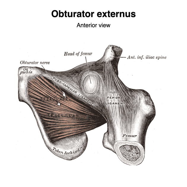
View Craig Hacking's current disclosuresThe obturator externus is a flat, triangular muscle, which covers the outer surface of the anterior wall of the pelvis.
Summary
Origin: external surface of obturator membrane and adjacent bone (inferior pubic ramus and the ramus of the ischium)
Insertion: trochanteric fossa of femur
Blood supply: obturator and medial circumflex femoral arteries
Innervation: Posterior division of obturator nerve (L3, L4)4
Action: thigh external rotation, stabilization of hip joint
Gross Anatomy
The obturator externus is a triangular muscle arising from the antero-lateral two thirds of the external surface of the obturator membrane. It passes laterally and posteriorly, beneath the neck of femur, narrowing into a tendon which inserts into the trochanteric fossa of the femur4.
The posterior aspect of the hip joint capsule extends along the neck of femur only as far as where the obturator externus tendon is in contact with the periosteum. While the anterior aspect of the joint capsule includes the whole neck of femur3.
The anterior division of the obturator nerve reaches the thigh by coursing in front of the obturator externus muscle belly. The posterior branch of the obturator nerve pierces the upper border of the muscle belly, giving off a branch to supply the muscle3,4.
Posterior to the muscle lie the obturator vessels, sandwiched between the muscle belly and obturator membrane3.
References
- 1. Gray's Anatomy for Students: With STUDENT CONSULT Online Access, 3e. Churchill Livingstone. ISBN:0702051314. Read it at Google Books - Find it at Amazon
- 2. Clinically Oriented Anatomy. Lippincott Williams & Wilkins. ISBN:1451119453. Read it at Google Books - Find it at Amazon
- 3. Last's Anatomy. Churchill Livingstone. ISBN:0702033944. Read it at Google Books - Find it at Amazon
- 4. Susan Standring. Gray's Anatomy. (2020) ISBN: 9780702077050 - Google Books
Incoming Links
Related articles: Anatomy: Lower limb
- skeleton of the lower limb
- joints of the lower limb
-
hip joint
- ligaments
- muscles
- additional structures
- hip joint capsule
- zona orbicularis
- iliotibial band
-
hip bursae
- anterior
- iliopsoas bursa (iliopectineal bursa)
- lateral
- subgluteal bursae
- greater trochanteric bursa (subgluteus maximus bursa)
- subgluteus medius bursa
- subgluteus minimus bursa
- gluteofemoral bursa
- subgluteal bursae
- postero-inferior
- anterior
- ossification centers
-
knee joint
- ligaments
- anterior cruciate ligament
- posterior cruciate ligament
- medial collateral ligament
- lateral collateral ligament
- meniscofemoral ligament (mnemonic)
-
posterolateral ligamentous complex
- arcuate ligament
- patellar tendon and quadriceps tendon
- anterolateral ligament
- posterior oblique ligament
- oblique popliteal ligament
- medial patellofemoral ligament
- additional structures
- extensor mechanism of the knee
- groove for the popliteus tendon
- knee bursae
- anterior bursae
- medial bursae
- lateral bursae
- posterior bursae
- knee capsule
- lateral patellar retinaculum
- medial patellar retinaculum
- menisci
- pes anserinus (mnemonic)
- ossification centers
- ligaments
- tibiofibular joints
-
ankle joint
- regional anatomy
- medial ankle
- lateral ankle
- anterior ankle
- ligaments
- medial collateral (deltoid) ligament
- lateral collateral ligament
- additional structures
- ankle bursae
- ossification centers of the ankle
- variants
- regional anatomy
- foot joints
- subtalar joint
- mid-tarsal (Chopart) joint
-
tarsometatarsal (Lisfranc) joint
- ligaments
- intermetatarsal joint
- metatarsophalangeal joint
- interphalangeal joint
- ossification centers
-
hip joint
- spaces of the lower limb
-
muscles of the lower limb
- muscles of the pelvic group
- muscles of the thigh
- muscles of the leg
- anterior compartment of the leg
- posterior compartments of the leg
- lateral compartment of the leg
- muscles of the foot
- dorsal muscles
- plantar muscles
- 1st layer
- 2nd layer
- 3rd layer
- 4th layer
- accessory muscles of the lower limb
- accessory gluteal muscles
-
accessory muscles of the ankle
- accessory peroneal muscles
- accessory flexor digitorum longus muscle
- accessory soleus muscle
- peroneocalcaneus internus muscle
- tibiocalcaneus internus muscle
- extensor hallucis capsularis tendon
- anterior fibulocalcaneus muscle
- accessory extensor digiti secundus muscle
- tibioastragalus anticus of Gruber muscle
- vascular supply of the lower limb
- arterial supply of the lower limb
- venous drainage of the lower limb
- innervation of the lower limb
- lymphatic system of the lower limb
- lymphatic pathways
- anteromedial group
- anterolateral group
- posteromedial group
- posterolateral group
- lower limb lymph nodes
- lymphatic pathways


 Unable to process the form. Check for errors and try again.