
Bipartite medial cuneiform
Citation, DOI, disclosures and article data
At the time the article was created Yuranga Weerakkody had no recorded disclosures.
View Yuranga Weerakkody's current disclosuresAt the time the article was last revised Arlene Campos had no financial relationships to ineligible companies to disclose.
View Arlene Campos's current disclosures- Bipartite medial cuneiform bone
- Bi-partite medial cuneiform
A bipartite medial cuneiform is an anatomical variant where there are two ossification centers involving the medial cuneiform. In many cases, the overall shape of the medial cuneiform is conserved, although the size of the two combined bones is larger than that of a normal medial cuneiform.
On this page:
Epidemiology
The estimated incidence is thought to be ~1% (range 0.3-2.4%) of the general population with the majority (~85%) reported in males 7.
Associations
Some patients may have additional associations which include:
- os intermetatarseum
- accessory navicular bone
- spinal segmentation anomalies
- lunotriquetral coalition
- an accessory temporal bone suture
- bipartite temporal bone
Clinical presentation
Most patients are asymptomatic although occasionally some patients can present with pain.
Gross anatomy
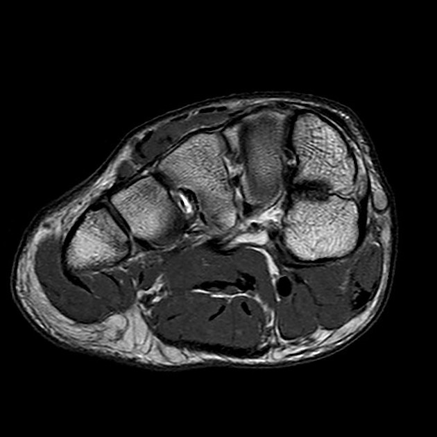
Bipartite medial cuneiforms are divided by an oblique/horizontal synchondrosis and/or syndesmosis into a smaller dorsal ossicle and larger plantar ossicle 7.
Radiographic features
Plain radiograph
A lateral foot radiograph (30° angled) may give an "E" sign with the two bones most often dorsal and plantar viewed end on.
MRI
May also give an "E" sign when viewed on sagittal images. MRI also allows direct visualization of anatomy as well as an appreciation of any signal abnormality along the synchondrosis.
Treatment and prognosis
Symptomatic cases may benefit from intervention ranging from various nonoperative measures to surgical excision.
See also
References
- 1. Brookes-Fazakerley S, Jackson G, Platt S. An Additional Middle Cuneiform? J Surg Case Rep. 2015;2015(7):rjv076. doi:10.1093/jscr/rjv076 - Pubmed
- 2. Chang GH, Chang EY, Chung CB et-al. Bipartite Medial Cuneiform: Case Report and Retrospective Review of 1000 Magnetic Resonance (MR) Imaging Studies. Case Rep Med. 2014;2014: 130979. doi:10.1155/2014/130979 - Free text at pubmed - Pubmed citation
- 3. Burnett SE, Case DT. Bipartite medial cuneiform: new frequencies from skeletal collections and a meta-analysis of previous cases. Homo. 2011;62 (2): 109-25. doi:10.1016/j.jchb.2011.01.002 - Pubmed citation
- 4. Chiodo CP, Parentis MA, Myerson MS. Symptomatic bipartite medial cuneiform in an adult athlete: a case report. Foot Ankle Int. 2002;23 (4): 348-51. Pubmed citation
- 5. Dellacorte MP, Lin PJ, Grisafi PJ. Bilateral bipartite medial cuneiform. A case report. J Am Podiatr Med Assoc. 1992;82 (9): 475-8. doi:10.7547/87507315-82-9-475 - Pubmed citation
- 6. O'Neal M, Ganey T, Ogden J. Fracture of a Bipartite Medial Cuneiform Synchondrosis. Foot Ankle Int. 1995;16(1):37-40. doi:10.1177/107110079501600108 - Pubmed
- 7. Serfaty et al. Bipartite Medial Cuneiform: Magnetic Resonance Imaging Findings and Prevalence of This Rare Anatomical Variant. (2020) Skeletal radiology. doi:10.1007/s00256-019-03353-3 - Pubmed
Incoming Links
Related articles: Anatomy: Lower limb
- skeleton of the lower limb
- joints of the lower limb
-
hip joint
- ligaments
- muscles
- additional structures
- hip joint capsule
- zona orbicularis
- iliotibial band
-
hip bursae
- anterior
- iliopsoas bursa (iliopectineal bursa)
- lateral
- subgluteal bursae
- greater trochanteric bursa (subgluteus maximus bursa)
- subgluteus medius bursa
- subgluteus minimus bursa
- gluteofemoral bursa
- subgluteal bursae
- postero-inferior
- anterior
- ossification centers
-
knee joint
- ligaments
- anterior cruciate ligament
- posterior cruciate ligament
- medial collateral ligament
- lateral collateral ligament
- meniscofemoral ligament (mnemonic)
-
posterolateral ligamentous complex
- arcuate ligament
- patellar tendon and quadriceps tendon
- anterolateral ligament
- posterior oblique ligament
- oblique popliteal ligament
- medial patellofemoral ligament
- additional structures
- extensor mechanism of the knee
- groove for the popliteus tendon
- knee bursae
- anterior bursae
- medial bursae
- lateral bursae
- posterior bursae
- knee capsule
- lateral patellar retinaculum
- medial patellar retinaculum
- menisci
- pes anserinus (mnemonic)
- ossification centers
- ligaments
- tibiofibular joints
-
ankle joint
- regional anatomy
- medial ankle
- lateral ankle
- anterior ankle
- ligaments
- medial collateral (deltoid) ligament
- lateral collateral ligament
- additional structures
- ankle bursae
- ossification centers of the ankle
- variants
- regional anatomy
- foot joints
- subtalar joint
- mid-tarsal (Chopart) joint
-
tarsometatarsal (Lisfranc) joint
- ligaments
- intermetatarsal joint
- metatarsophalangeal joint
- interphalangeal joint
- ossification centers
-
hip joint
- spaces of the lower limb
-
muscles of the lower limb
- muscles of the pelvic group
- muscles of the thigh
- muscles of the leg
- anterior compartment of the leg
- posterior compartments of the leg
- lateral compartment of the leg
- muscles of the foot
- dorsal muscles
- plantar muscles
- 1st layer
- 2nd layer
- 3rd layer
- 4th layer
- accessory muscles of the lower limb
- accessory gluteal muscles
-
accessory muscles of the ankle
- accessory peroneal muscles
- accessory flexor digitorum longus muscle
- accessory soleus muscle
- peroneocalcaneus internus muscle
- tibiocalcaneus internus muscle
- extensor hallucis capsularis tendon
- anterior fibulocalcaneus muscle
- accessory extensor digiti secundus muscle
- tibioastragalus anticus of Gruber muscle
- vascular supply of the lower limb
- arterial supply of the lower limb
- venous drainage of the lower limb
- innervation of the lower limb
- lymphatic system of the lower limb
- lymphatic pathways
- anteromedial group
- anterolateral group
- posteromedial group
- posterolateral group
- lower limb lymph nodes
- lymphatic pathways


 Unable to process the form. Check for errors and try again.