
The knee joint is a modified hinge joint between the femur, tibia, and patella. It is the largest synovial joint in the body and allows flexion and extension of the leg as well as some rotation in the flexed position.
On this page:
Summary
location: two condylar joints between femur and tibia; saddle joint between patella and femur
blood supply: main supply are the genicular branches of the popliteal artery
nerve supply: branches from the femoral, tibial, common peroneal, and obturator nerves
movement: flexion to 150°, extension to 5-10° hyperextension; rotation whilst in the flexed position to 10° actively and 60° passively
Gross anatomy
Articulations
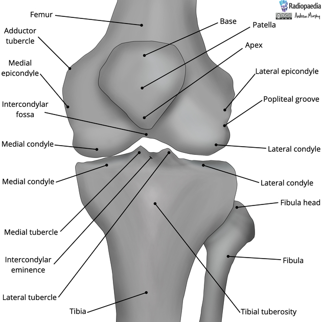
There are two condylar joints between the femur and tibia (tibiofemoral). There are medial and lateral articular facets on the tibial plateau and medial and lateral femoral condyles on the distal femur which are convex and circular shaped.
medially: between a narrow and curved femoral condyle, and an oval tibial articular surface with a long anteroposterior length
laterally: between a wide and flat femoral condyle; and a circular tibial articular surface which overhangs the shaft posterolaterally
the knee menisci are shaped accordingly
Saddle joint between the patella and femoral condyles:
medial, lateral and odd facet on the posterior surface of the patella articulate with the medial and lateral condyles of the femur
on flexion, more parts of the bony surface are exposed to articulation (four below, odd facet) and are more proximal on the patella
with extension, the contact area lessens and moves distally
Menisci
fibrocartilaginous, C-shaped in appearance and triangular in cross-section
the medial meniscus is attached to the medial collateral ligament and the lateral meniscus is attached to the popliteus tendon
attached to the femur and tibia via the coronary ligaments
See menisci of the knee.
Joint capsule
-
knee capsule
-
on the femur
-
adheres below the epiphyseal line down to the articular margin except in two places
posteriorly attached to the intercondylar ridge at the lower limit of the popliteal surface
on the lateral condyle it encloses a pit and groove for the popliteus tendon
-
-
on the tibia
-
attached around the margins of the tibial plateau except in two places
posteriorly to the ridge between the two condyles at the lower end of the groove for the PCL
laterally the capsule is not attached to the tibia but is prolonged down over the popliteus tendon
-
-
two main gaps
lateroposteriorly allowing passage of the popliteus tendon 1
anteriorly a circular gap whose margins attach to the patella allowing communication with suprapatellar bursa 1
-
Synovial membrane
joint capsule is lined by synovial membrane, however, the attachment of the synovial membrane does not coincide with the capsular attachments because of the intra-articular structures
the cruciate ligament and popliteus tendon are extrasynovial but intracapsular
communicates with the suprapatellar bursa
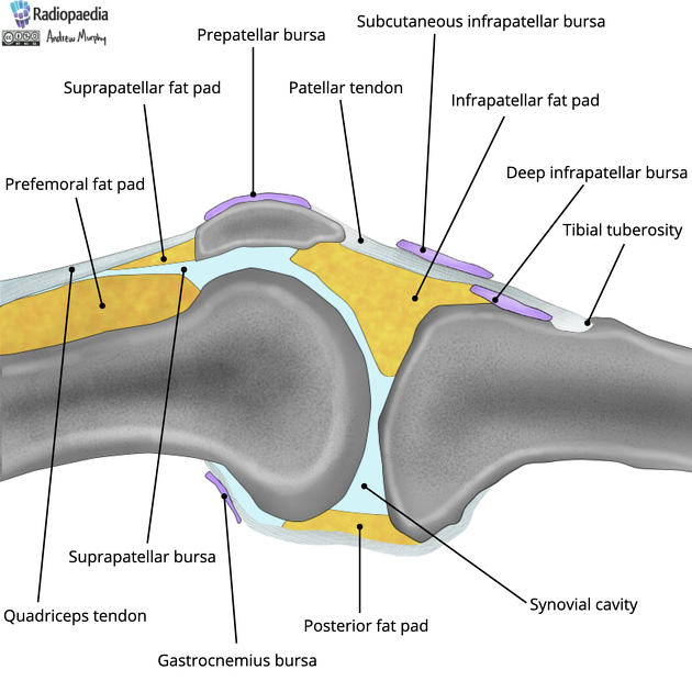
Fat pads
There are three anterior fat pads:
Attachments
-
intracapsular ligaments
-
anterior intermeniscal ligament
connects the anterior limbs of the two menisci
-
anterior (Humphrey) and posterior (Wrisberg) meniscofemoral ligaments:
the lateral meniscus is attached to the medial femoral condyle via the anterior and posterior meniscofemoral ligament of Humphrey and Wrisberg
-
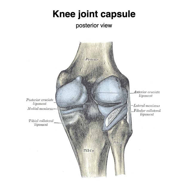
cruciate ligaments: cross each other to form an "x" shape.
anterior cruciate ligament: from the anterior tibial plateau to the lateral femoral condyle
posterior cruciate ligament: from the posterior intercondylar area to the medial femoral condyle
-
-
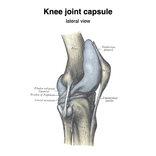
extracapsular ligaments
patellar retinacular ligaments: medial and lateral portions of the quadriceps tendon pass down on either side of the patella and are inserted into the upper extremity of the tibia on either side of the tuberosity, merging into the capsule
-
from the medial epicondyle to the medial surface of the tibia, which it is separated from by the passage of the inferior medial genicular arteries
attached to the medial meniscus
flat band like approximately 12 cm long
has superficial and deep parts (thickening of the capsule)
-
from the lateral epicondyle to the fibular head
not attached to the lateral meniscus
thin cord like, approximately 5 cm long
separated from the tibia within the joint by the popliteus tendon and outside the joint by the inferior lateral genicular artery
-
tendinous expansion of the semimembranosus muscle terminating on the popliteal surface of the femur
perforated by the middle genicular artery
-
thickened part of the joint capsule that arches over the popliteus tendon as it emerges from the joint capsule and attached to the styloid process of the fibular head
-
extends from the popliteus tendon near the myotendinous junction to the posterior aspect of the fibular styloid process, posteromedial to the biceps insertion
-
from the apex of the patella to the tibial tuberosity
-
other
-
tendons
Bursa
suprapatellar - superior extension of the knee joint cavity
prepatellar - communicates with the joint cavity, between the lower half of the patella and skin
subcutaneous infrapatellar - between the patella ligament and skin
deep infrapatellar - between the tibia and patella tendon
-
posterior (between muscle and bone)
popliteal - communicates with the joint cavity, beneath the tendon of popliteus lying in the gutter between tibia and head of fibula
-
gastrocnemius
bursa beneath the medial head (and usually the lateral head) communicates with the joint cavity
semimembranosus - may communicate with the bursa beneath the medial head of the gastrocnemius
Relations
Blood supply
The knee is supplied by anastomoses of:
-
five genicular branches of the popliteal artery (main supply)
medial and lateral superior genicular arteries encircle the femoral condyle
medial and lateral inferior genicular arteries encircle the tibial condyle
middle genicular artery supplies the anterior and posterior cruciate ligaments
circumflex fibular branches of the posterior tibial artery
anterior and posterior recurrent branches of the anterior tibial artery
Innervation
Multiple articular branches are derived from several nerves (Hilton's law):
branches of the femoral nerve to vastus medialis, and also intermedius and lateralis
from the sciatic nerve by genicular branches of the tibial and common peroneal nerves
from the obturator nerve by a branch from the posterior division
Movements
-
flexion
semimembranosus, semitendinosus, biceps femoris, gracilis, sartorius
also gastrocnemius, plantaris and popliteus
-
extension
quadriceps femoris, iliotibial tract
also gluteus maximus, tensor fascia latae
-
internal rotation (when flexed)
semimembranosus, semitendinosus, gracilis, sartorius
-
external rotation (when flexed)
biceps femoris
-
unlocking
popliteus externally rotates femur on tibia, locked ligaments loosen, hamstrings can then flex free
-
locking
as the knee moves into full extension, the anterior cruciate ligament becomes taut, with no further extension of the lateral condyle possible
passive rotation forwards of the lateral condyle around the radius of the taut anterior cruciate ligament
medial femoral condyle is then able to glide backwards into full extension
tightening of the oblique popliteal, lateral collateral and medial collateral ligaments
purely passive due to the skew pull of the obliquely set ligaments
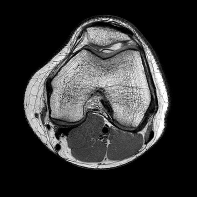
Radiographic features
Plain radiograph
See knee radiograph (an approach)
Related pathology
Stieda fracture (MCL avulsion fracture)


 Unable to process the form. Check for errors and try again.