
Lateral pterygoid muscle
Citation, DOI, disclosures and article data
At the time the article was created Frank Gaillard had no recorded disclosures.
View Frank Gaillard's current disclosuresAt the time the article was last revised Tea Elliott had no financial relationships to ineligible companies to disclose.
View Tea Elliott's current disclosures- Lateral pterygoid
- Pterygoideus externus
- External pterygoid muscle
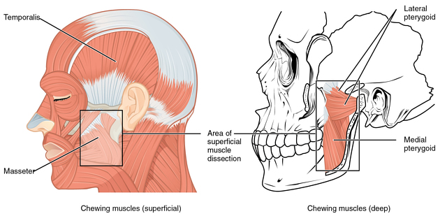
The lateral pterygoid muscle, also known as pterygoideus externus or external pterygoid muscle, is one of the muscles of mastication.
On this page:
Summary
origin: superior head from the infratemporal surface and the infratemporal crest of the greater wing of the sphenoid bone. Inferior head from lateral surface of the lateral pterygoid plate
insertion: pterygoid fovea on the anterior neck of the mandible
innervation: nerve to lateral pterygoid, a branch of the anterior division of the mandibular division of the trigeminal nerve
action: opening of the mouth, retrusion of the temperomandibular joint, side-to-side movement of the jaw 1
Gross anatomy
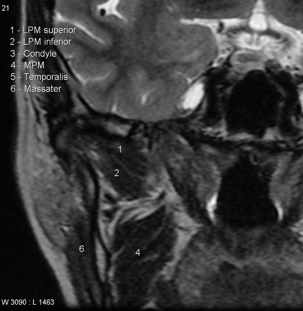
The lateral pterygoid is a short and thick muscle with a somewhat conical form. It extends almost horizontally, posteriorly, and laterally between the infratemporal fossa and the condyle of the mandible. It has two heads: an upper (superior) and a lower (inferior).
The superior part arises from the lower part of the lateral surface of the greater wing of the sphenoid bone and the infratemporal crest. It inserts into the temporomandibular joint capsule and the temporomandibular disc.
The inferior part arises from the lateral surface of the lateral pterygoid plate and inserts into a depression in front of the neck of the condyle of the mandible, the pterygoid fovea 1.
ADVERTISEMENT: Supporters see fewer/no ads
Innervation
The muscle is supplied by the paired nerves to lateral pterygoid (one for each head), which arise deep to the muscle from the anterior division of the mandibular nerve (CN V3).
Action
The superior part is active during retrusion (opposite of protrusion) and ipsilateral jaw movement. It is also essential in pulling the capsule and disc forward during mouth opening, thereby maintaining normal relationship between the condyle of the mandible and the disc of the tempoeromandibular joint.
The inferior part is responsible for opening of the mouth, protrusion, and contralateral jaw movement.
Hyperactivity of the lateral pterygoid muscle has been described in temperomandibular joint internal derangement, especially with longstanding anterior displacement of the disc without recapture. Thickening of the tendon (inferior part) can give rise to the "double disc sign".
Variant anatomy
Anatomical variants of the lateral pterygoid include:
fusion with temporalis or digastric muscle
-
variation in number of heads
three headed variant with an inner head originating at the greater wing of the sphenoid
single headed variant
-
variation in number of insertions
three insertion sites: articular disc, TMJ capsule, condyle of the mandible
single insertion site at the condyle only
References
- 1. Susan Standring. Gray's Anatomy. (2020) ISBN: 9780702077050 - Google Books
- 2. Anatomy of the Human Body. (2000) ISBN: 1587341026 - Google Books
- 3. Sommer O, Aigner F, Rudisch A et al. Cross-Sectional and Functional Imaging of the Temporomandibular Joint: Radiology, Pathology, and Basic Biomechanics of the Jaw. Radiographics. 2003;23(6):e14. doi:10.1148/rg.e14 - Pubmed
- 4. Tomas X, Pomes J, Berenguer J et al. MR Imaging of Temporomandibular Joint Dysfunction: A Pictorial Review. Radiographics. 2006;26(3):765-81. doi:10.1148/rg.263055091 - Pubmed
- 5. R. Shane Tubbs, Mohammadali M. Shoja, Marios Loukas. Bergman's Comprehensive Encyclopedia of Human Anatomic Variation. (2016) ISBN: 9781118430354 - Google Books
Incoming Links
- Deep temporal nerves
- Infratemporal fossa
- Greater wing of sphenoid
- Muscles of mastication
- Motor nucleus of the trigeminal nerve
- Masticatory muscle hypertrophy
- Accessory meningeal artery
- Condylar process of the mandible
- Temporomandibular joint disc
- Maxillary artery
- Oropharyngeal (p16-negative) cancer (staging)
- Temporomandibular joint
- Medial pterygoid muscle
- Pterygomandibular space
- Lingual nerve
- Inferior alveolar nerve
- Masseteric nerve
- Pterygoid processes
- Trigeminal nerve
- Pterygoid fovea
Related articles: Anatomy: Head and neck
- skeleton of the head and neck
-
cranial vault
- scalp (mnemonic)
- fontanelle
-
sutures
- calvarial
- facial
- frontozygomatic suture
- frontomaxillary suture
- frontolacrimal suture
- frontonasal suture
- temporozygomatic suture
- zygomaticomaxillary suture
- parietotemporal suture (parietomastoid suture)
- occipitotemporal suture (occipitomastoid suture)
- sphenofrontal suture
- sphenozygomatic suture
- spheno-occipital suture (not a true suture)
- lacrimomaxillary suture
- nasomaxillary suture
- internasal suture
- basal/internal
- skull landmarks
- frontal bone
- temporal bone
- parietal bone
- occipital bone
- skull base (foramina)
-
facial bones
- midline single bones
- paired bilateral bones
- cervical spine
- hyoid bone
- laryngeal cartilages
-
cranial vault
- muscles of the head and neck
- muscles of the tongue (mnemonic)
- muscles of mastication
-
facial muscles
- epicranius muscle
- circumorbital and palpebral muscles
- nasal muscles
-
buccolabial muscles
- elevators, retractors and evertors of the upper lip
- levator labii superioris alaeque nasalis muscle
- levator labii superioris muscle
- zygomaticus major muscle
- zygomaticus minor muscle
- levator anguli oris muscle
- malaris muscle
- risorius muscle
- depressors, retractors and evertors of the lower lip
- depressor labii inferioris muscle
- depressor anguli oris muscle
- mentalis muscle
- compound sphincter
-
orbicularis oris muscle
- incisivus labii superioris muscle
- incisivus labii inferioris muscle
-
orbicularis oris muscle
- muscle of mastication
- modiolus
- elevators, retractors and evertors of the upper lip
- muscles of the middle ear
- orbital muscles
- muscles of the soft palate
- pharyngeal muscles
- suprahyoid muscles
- infrahyoid muscles
- intrinsic muscles of the larynx
- muscles of the neck
- platysma muscle
- longus colli muscle
- longus capitis muscle
- scalenus anterior muscle
- scalenus medius muscle
- scalenus posterior muscle
- scalenus pleuralis muscle
- sternocleidomastoid muscle
-
suboccipital muscles
- rectus capitis posterior major muscle
- rectus capitis posterior minor muscle
- obliquus capitis superior muscle
- obliquus capitis inferior muscle
- accessory muscles of the neck
- deep cervical fascia
-
deep spaces of the neck
- anterior cervical space
- buccal space
- carotid space
- danger space
- deep cervical fascia
- infratemporal fossa
- masticator space
- parapharyngeal space
- stylomandibular tunnel
- parotid space
- pharyngeal (superficial) mucosal space
- perivertebral space
- posterior cervical space
- pterygopalatine fossa
- retropharyngeal space
- suprasternal space (of Burns)
- visceral space
- surgical triangles of the neck
- orbit
- ear
- paranasal sinuses
- upper respiratory tract
- viscera of the neck
- blood supply of the head and neck
-
arterial supply
-
common carotid artery
- carotid body
- carotid bifurcation
- subclavian artery
- variants
-
common carotid artery
- venous drainage
-
arterial supply
- innervation of the head and neck
-
cranial nerves
- olfactory nerve (CN I)
- optic nerve (CN II)
- oculomotor nerve (CN III)
- trochlear nerve (CN IV)
-
trigeminal nerve (CN V) (mnemonic)
- trigeminal ganglion
- ophthalmic division
- maxillary division
- mandibular division
- abducens nerve (CN VI)
- facial nerve (CN VII)
-
vestibulocochlear nerve (CN VIII)
- vestibular ganglion (Scarpa's ganglion)
- glossopharyngeal nerve (CN IX)
- vagus nerve (CN X)
- (spinal) accessory nerve (CN XI)
- hypoglossal nerve (CN XII)
- parasympathetic ganglia of the head and neck
- cervical sympathetic ganglia
- greater occipital nerve
- third occipital nerve
-
cervical plexus
- muscular branches
- longus capitis
- longus colli
- scalenes
- geniohyoid
- thyrohyoid
-
ansa cervicalis
- omohyoid (superior and inferior bellies separately)
- sternothyroid
- sternohyoid
- phrenic nerve
- contribution to the accessory nerve (CN XI)
- cutaneous branches
- muscular branches
- brachial plexus
- pharyngeal plexus
-
cranial nerves
- lymphatic drainage of the head and neck
- embryological development of the head and neck


 Unable to process the form. Check for errors and try again.