
Ligamentum mucosum
Citation, DOI, disclosures and article data
At the time the article was created Amir Rezaee had no recorded disclosures.
View Amir Rezaee's current disclosuresAt the time the article was last revised Henry Knipe had the following disclosures:
- Radiopaedia Events Pty Ltd, Speaker fees (past)
- Integral Diagnostics, Shareholder (ongoing)
- Micro-X Ltd, Shareholder (ongoing)
These were assessed during peer review and were determined to not be relevant to the changes that were made.
View Henry Knipe's current disclosures- Infrapatellar plica
The ligamentum mucosum, also known as the infrapatellar plica, is an embryological remnant of a synovial septum and one of the types of knee plicae.
On this page:
Epidemiology
Ligamentum mucosum is the most common knee plica, with an incidence of ~75% (range 65%-86%) 4,5.
Classification
The classification according to the plica morphology was proposed by Kim 4.
separate type: completely separated from the ACL (60.5%)
split type: longitudinally divided (13.5%)
vertical septum type: attached to the ACL (10.5%)
fenestra type: with fenestrated vertical septum (1.0%)
Radiographic features
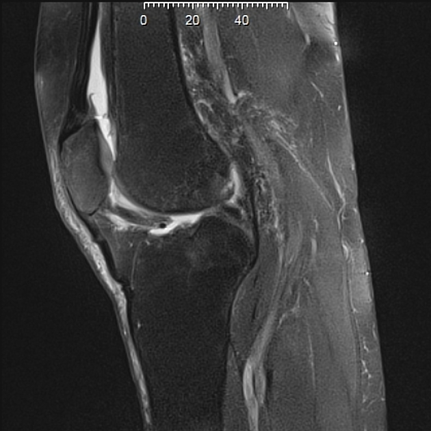
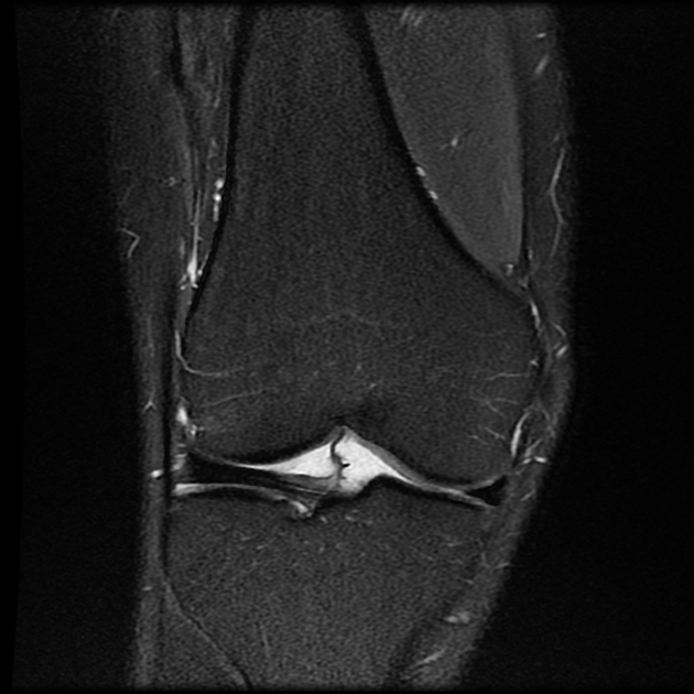
MRI
The ligamentum mucosum is best seen on T2/PD sagittal images as a curvilinear, low signal structure, originating from the anterior intercondylar notch and traversing the infrapatellar (Hoffa's) fat pad to attach to the inferior pole of the patella. It may connect to the anterior cruciate ligament (ACL, the anterior transverse meniscomeniscal ligament, or anterior horn of the lateral meniscus 1. Its thickness ranges from very thin to almost as thick as the anterior cruciate ligament.
Related pathology
References
- 1. Cothran R, McGuire P, Helms C, Major N, Attarian D. MR Imaging of Infrapatellar Plica Injury. AJR Am J Roentgenol. 2003;180(5):1443-7. doi:10.2214/ajr.180.5.1801443 - Pubmed
- 2. García-Valtuille R, Abascal F, Cerezal L et al. Anatomy and MR Imaging Appearances of Synovial Plicae of the Knee. Radiographics. 2002;22(4):775-84. doi:10.1148/radiographics.22.4.g02jl03775 - Pubmed
- 3. Cothran R, McGuire P, Helms C, Major N, Attarian D. MR Imaging of Infrapatellar Plica Injury. AJR Am J Roentgenol. 2003;180(5):1443-7. doi:10.2214/ajr.180.5.1801443 - Pubmed
- 4. Kosarek F & Helms C. The MR Appearance of the Infrapatellar Plica. AJR Am J Roentgenol. 1999;172(2):481-4. doi:10.2214/ajr.172.2.9930807 - Pubmed
- 5. Kim S, Min B, Kim H. Arthroscopic Anatomy of the Infrapatellar Plica. Arthroscopy. 1996;12(5):561-4. doi:10.1016/s0749-8063(96)90195-4 - Pubmed
Incoming Links
Related articles: Anatomy: Lower limb
- skeleton of the lower limb
- joints of the lower limb
-
hip joint
- ligaments
- muscles
- additional structures
- hip joint capsule
- zona orbicularis
- iliotibial band
-
hip bursae
- anterior
- iliopsoas bursa (iliopectineal bursa)
- lateral
- subgluteal bursae
- greater trochanteric bursa (subgluteus maximus bursa)
- subgluteus medius bursa
- subgluteus minimus bursa
- gluteofemoral bursa
- subgluteal bursae
- postero-inferior
- anterior
- ossification centers
-
knee joint
- ligaments
- anterior cruciate ligament
- posterior cruciate ligament
- medial collateral ligament
- lateral collateral ligament
- meniscofemoral ligament (mnemonic)
-
posterolateral ligamentous complex
- arcuate ligament
- patellar tendon and quadriceps tendon
- anterolateral ligament
- posterior oblique ligament
- oblique popliteal ligament
- medial patellofemoral ligament
- additional structures
- extensor mechanism of the knee
- groove for the popliteus tendon
- knee bursae
- anterior bursae
- medial bursae
- lateral bursae
- posterior bursae
- knee capsule
- lateral patellar retinaculum
- medial patellar retinaculum
- menisci
- pes anserinus (mnemonic)
- ossification centers
- ligaments
- tibiofibular joints
-
ankle joint
- regional anatomy
- medial ankle
- lateral ankle
- anterior ankle
- ligaments
- medial collateral (deltoid) ligament
- lateral collateral ligament
- additional structures
- ankle bursae
- ossification centers of the ankle
- variants
- regional anatomy
- foot joints
- subtalar joint
- mid-tarsal (Chopart) joint
-
tarsometatarsal (Lisfranc) joint
- ligaments
- intermetatarsal joint
- metatarsophalangeal joint
- interphalangeal joint
- ossification centers
-
hip joint
- spaces of the lower limb
-
muscles of the lower limb
- muscles of the pelvic group
- muscles of the thigh
- muscles of the leg
- anterior compartment of the leg
- posterior compartments of the leg
- lateral compartment of the leg
- muscles of the foot
- dorsal muscles
- plantar muscles
- 1st layer
- 2nd layer
- 3rd layer
- 4th layer
- accessory muscles of the lower limb
- accessory gluteal muscles
-
accessory muscles of the ankle
- accessory peroneal muscles
- accessory flexor digitorum longus muscle
- accessory soleus muscle
- peroneocalcaneus internus muscle
- tibiocalcaneus internus muscle
- extensor hallucis capsularis tendon
- anterior fibulocalcaneus muscle
- accessory extensor digiti secundus muscle
- tibioastragalus anticus of Gruber muscle
- vascular supply of the lower limb
- arterial supply of the lower limb
- venous drainage of the lower limb
- innervation of the lower limb
- lymphatic system of the lower limb
- lymphatic pathways
- anteromedial group
- anterolateral group
- posteromedial group
- posterolateral group
- lower limb lymph nodes
- lymphatic pathways


 Unable to process the form. Check for errors and try again.