
Mucinous cystadenoma of the pancreas
Citation, DOI, disclosures and article data
At the time the article was created Yuranga Weerakkody had no recorded disclosures.
View Yuranga Weerakkody's current disclosuresAt the time the article was last revised Mohammad Taghi Niknejad had no financial relationships to ineligible companies to disclose.
View Mohammad Taghi Niknejad's current disclosures- Mucinous cystadenoma of pancreas
- Pancreatic mucinous cystadenoma
- MCN pancreas
- Mucinous cystadenoma (MCN) of pancreas
- Mucinous cystadenoma (pancreas)
Mucinous cystadenomas (MCN) of the pancreas are a type of mucinous cystic neoplasm of the pancreas.
On this page:
Epidemiology
Previously believed to occur exclusively in middle age females 5, it has occasionally been described in males 6,7.
Pathology
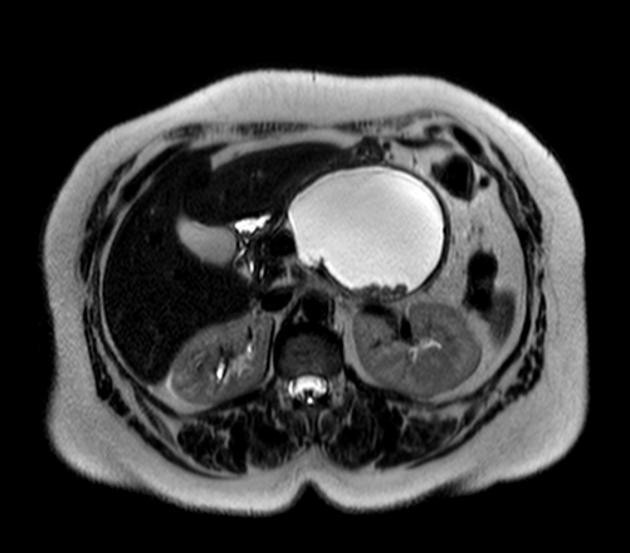
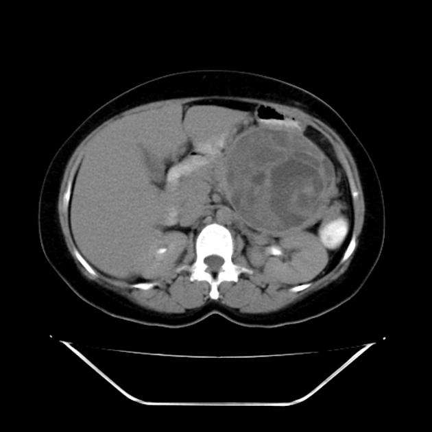
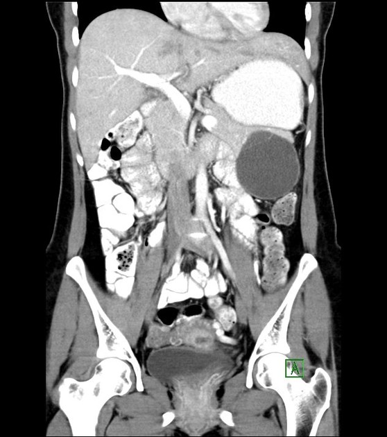
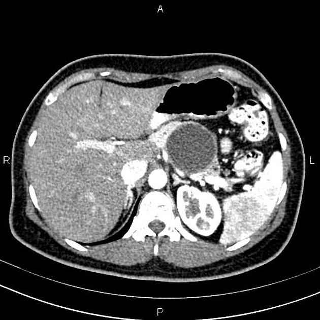
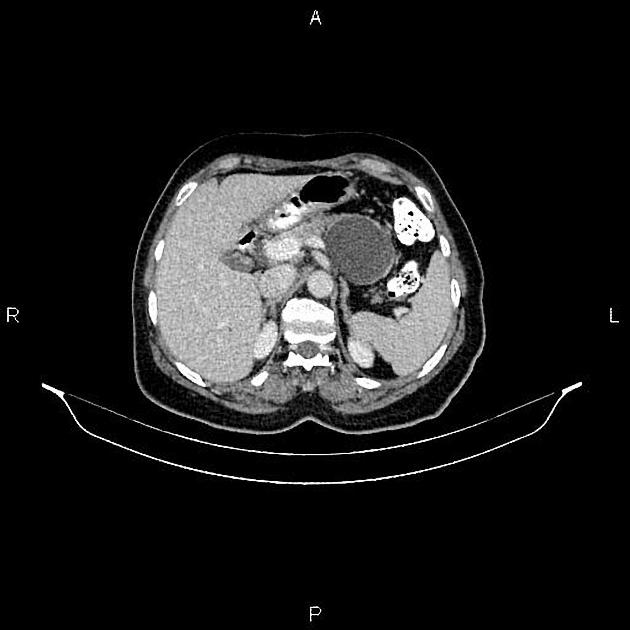
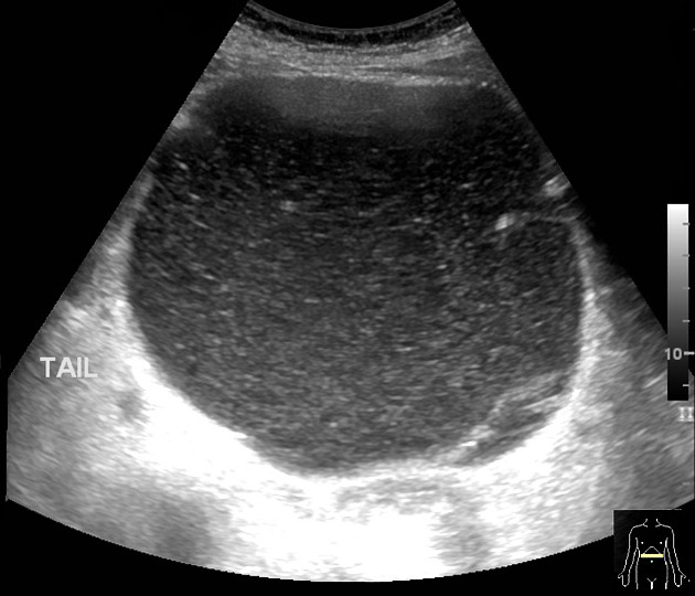
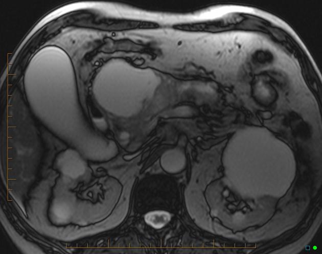
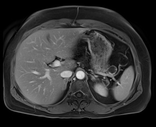
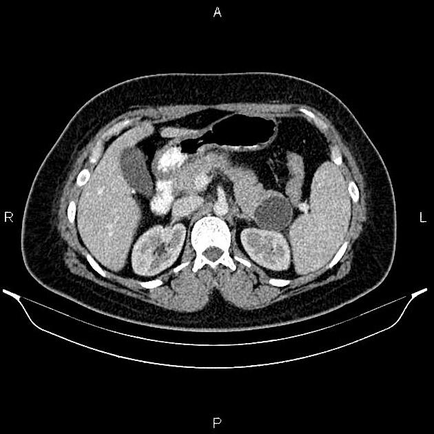
It is a large uni/multilocular cystic pancreatic neoplasm lined by columnar mucinous epithelium. While mucinous cystadenomas very infrequently communicate with the pancreatic duct 13, they can cause partial pancreatic ductal obstruction 11. They are considered premalignant or malignant lesions with usually elevated CEA and CA 19-9 serum levels.
Location
Largely (~80%) occur in the body or tail of the pancreas, and less commonly in the head of the pancreas (~20%) 11.
Radiographic features
CT
- the tumor contour tends to be rounded or ovoid although this is not an absolutely specific feature 2
- associated calcification when present tends to be more peripheral 1,11
- contents of the lesion may be heterogenous in attenuation 2
- internal septations may be present and tend to be linear or curvilinear 2
Differential diagnosis
On ultrasound or CT consider:
- mucinous cystadenocarcinoma of the pancreas: at times almost impossible to differentiate on ultrasound or CT from a mucinous cystadenoma 8
- pancreatic pseudocyst
- oligocystic variant of serous cystadenoma of the pancreas
- pancreatic hydatid cyst 14
References
- 1. Curry CA, Eng J, Horton KM et-al. CT of primary cystic pancreatic neoplasms: can CT be used for patient triage and treatment? AJR Am J Roentgenol. 2000;175 (1): 99-103. AJR Am J Roentgenol (full text) - Pubmed citation
- 2. Lv P, Mahyoub R, Lin X et-al. Differentiating pancreatic ductal adenocarcinoma from pancreatic serous cystadenoma, mucinous cystadenoma, and a pseudocyst with detailed analysis of cystic features on CT scans: a preliminary study. Korean J Radiol. 12 (2): 187-95. doi:10.3348/kjr.2011.12.2.187 - Free text at pubmed - Pubmed citation
- 3. Wouters K, Ectors N, Van steenbergen W et-al. A pancreatic mucinous cystadenoma in a man with mesenchymal stroma, expressing oestrogen and progesterone receptors. Virchows Arch. 1998;432 (2): 187-9. Virchows Arch. (link) - Pubmed citation
- 4. Tajiri T, Tate G, Inagaki T et-al. Mucinous cystadenoma of the pancreas 17 years after excision of gallbladder because of a choledochal cyst. J. Gastroenterol. 2004;39 (2): 181-7. doi:10.1007/s00535-003-1271-z - Pubmed citation
- 5. Lee WA. Mucinous cystadenoma of the pancreas with predominant stroma creating a solid tumor. World J Surg Oncol. 2005;3 : 59. doi:10.1186/1477-7819-3-59 - Free text at pubmed - Pubmed citation
- 6. Suzuki M, Fujita N, Onodera H et-al. Mucinous cystic neoplasm in a young male patient. J. Gastroenterol. 2005;40 (11): 1070-4. doi:10.1007/s00535-005-1697-6 - Pubmed citation
- 7. Tokuyama Y, Osada S, Sanada Y et-al. Mucinous cystic neoplasm of the pancreas in a male patient. Rare Tumors. 2011;3 (2): e14. doi:10.4081/rt.2011.e14 - Free text at pubmed - Pubmed citation
- 8. Mathieu D, Guigui B, Valette PJ et-al. Pancreatic cystic neoplasms. Radiol. Clin. North Am. 1989;27 (1): 163-76. - Pubmed citation
- 9. Itai Y, Moss AA, Ohtomo K. Computed tomography of cystadenoma and cystadenocarcinoma of the pancreas. Radiology. 1982;145 (2): 419-25. Radiology (abstract) - Pubmed citation
- 10. Sahani DV, Kadavigere R, Saokar A et-al. Cystic pancreatic lesions: a simple imaging-based classification system for guiding management. Radiographics. 25 (6): 1471-84. doi:10.1148/rg.256045161 - Pubmed citation
- 11. Gore RM, Wenzke DR, Thakrar KH et-al. The incidental cystic pancreas mass: a practical approach. Cancer Imaging. 2012;12 (2): 414-21. doi:10.1102/1470-7330.2012.9054 - Free text at pubmed - Pubmed citation
- 12. Park JW, Jang JY, Kang MJ et-al. Mucinous cystic neoplasm of the pancreas: is surgical resection recommended for all surgically fit patients?. Pancreatology. 2014;14 (2): 131-6. doi:10.1016/j.pan.2013.12.006 - Pubmed citation
- 13. Morel A, Marteau V, Chambon E et-al. Pancreatic mucinous cystadenoma communicating with the main pancreatic duct on MRI. Br J Radiol. 2009;82 (984): e243-5. doi:10.1259/bjr/98185084 - Free text at pubmed - Pubmed citation
- 14. Varun D, Venkatarami Reddy V, Sivaramakrishna G, Chandramaliteeswaran C, Brahmeswara Rao M. Pancreatic hydatid cyst mimicking mucinous cystic neoplasm of pancreas: A case report. (2016) HPB. 18: e341. doi:10.1016/j.hpb.2016.02.883
Incoming Links
- Pancreatic mucinous cystadenoma
- Mucinous cystadenoma of the pancreas
- Pancreatic mucinous cystadenoma
- Mucinous cystadenoma of the pancreas
- Mucinous cystic lesion of the pancreas
- Serous cystadenoma of the pancreas - macrocystic/oligocystic
- Mucinous cystic neoplasm of the pancreas
- Mucinous cystic neoplasm of the pancreas
- Pancreatic mucinous cystadenoma
Related articles: Pathology: Hepato-Pancreato-Biliary
- liver
- depositional disorders
- infection and inflammation
- liver abscess
- hepatic hydatid infection
- cirrhosis
- hepatitis
- cholecystitis
- cholangitis
- malignancy
- liver and intrahepatic bile duct tumors
- benign epithelial tumors
- hepatocellular hyperplasia
- hepatocellular adenoma
- hepatic/biliary cysts
- benign nonepithelial tumors
- primary malignant epithelial tumors
- hepatocellular carcinoma
- hepatocellular carcinoma variants
-
cholangiocarcinoma
- intra-hepatic
- mass-forming type
- periductal infiltrating type - Klatskin tumors
- intraductal growing type
- extra-hepatic/large duct type
- intra-hepatic
- biliary cystadenocarcinoma
- combined hepatocellular and cholangiocarcinoma
- hepatoblastoma
- undifferentiated carcinoma
- primary malignant nonepithelial tumors
- hematopoietic and lymphoid tumors
- primary hepatic lymphoma
- hepatic myeloid sarcoma (hepatic chloroma)
- secondary tumors
- miscellaneous
- adrenal rest tumors
- hepatic carcinosarcoma
- hepatic fibroma
- hepatic hemangioma
- hepatic Kaposi sarcoma
- hepatic lipoma
- hepatic mesenchymal hamartoma
- hepatic myxoma
- hepatic rhabdoid tumor
- hepatic solitary fibrous tumor
- hepatic teratoma
- hepatic yolk sac tumor
- inflammatory myofibroblastic tumor (inflammatory pseudotumor)
- nodular regenerative hyperplasia
- pancreatic rest tumors
- primary hepatic carcinoid
- benign epithelial tumors
- liver and intrahepatic bile duct tumors
- metabolic
- trauma
-
vascular and perfusion disorders
- portal vein related
- hepatic artery related
- hepatic veins related
- inferior vena cava related
- other
- third inflow
- liver thrombotic angiitis
- infra diaphragmatic total anomalous pulmonary venous return (TAPVR)
- hereditary hemorrhagic telangiectasia (Osler-Weber-Rendu disease)
- pancreas
-
pancreatic neoplasms
- cystic neoplasm (cystic pancreatic mass differential diagnosis)
- solid neoplasm
- non-epithelial pancreatic neoplasms
- others
- simple pancreatic cyst
-
pancreatitis (mnemonic for the causes)
- acute pancreatitis
- chronic pancreatitis
- Ascaris-induced pancreatitis
- tropical pancreatitis
- autoimmune pancreatitis
- emphysematous pancreatitis
- hypertriglyceridemia-induced pancreatitis
- hereditary pancreatitis
- pancreatitis associated with cystic fibrosis
- pancreaticopleural fistula
- segmental pancreatitis
- pancreatic atrophy
- pancreatic lipomatosis
- pancreatic trauma
- pancreatic transplant
-
pancreatic neoplasms
- gallbladder and biliary
- congenital malformations and anatomical variants
- gallstones
- gallbladder inflammation
- bile ducts inflammation
- gallbladder wall abnormalities
- other gallbladder abnormalities
- bile duct dilatation (differential)
- bile duct wall thickening (differential)
- bile ducts neoplasms


 Unable to process the form. Check for errors and try again.