
Perinephric bridging septa
Citation, DOI, disclosures and article data
At the time the article was created Pradosh Kumar Sarangi had no recorded disclosures.
View Pradosh Kumar Sarangi's current disclosuresAt the time the article was last revised Daniel J Bell had no financial relationships to ineligible companies to disclose.
View Daniel J Bell's current disclosures- Kunin’s septa
- Kunin septa
- Septa of Kunin
- Perinephric bridging septum
- Septum of Kunin
- Kunin septum
- Kunin's septum
Perinephric bridging septa or septa of Kunin (singular: septum) are composed of numerous fibrous lamellae which traverse the perinephric fat 1,2 where they suspend the kidneys within the perirenal space. The septa may act as a barrier or conduit for the spread of pus, blood, urine, and neoplasms in the perinephric space.
In traumatic bleeding, the pressures generated in these closed spaces are considerable and may be adequate to tamponade a bleeding point, which may lead to conservative management.
On this page:
Gross anatomy
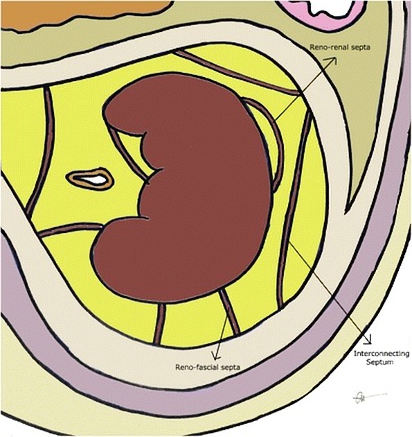
Three types of bridging septa have been described:
renorenal: septa running parallel to the renal capsule and attaching back onto the kidney
renofascial: septa connecting the capsule to the adjacent anterior or posterior renal fascia
interconnecting fascia: connecting the anterior and posterior layers of the perinephric fascia
Radiographic features
The septa can be appreciated as fine lines within the perinephric space extending from the surface of the kidney on CT and MRI.
History and etymology
The septa were first described by Milton Kunin, an American uroradiologist 1.
References
- 1. Kunin M. Bridging septa of the perinephric space: anatomic, pathologic, and diagnostic considerations. (1986) Radiology. 158 (2): 361-5. doi:10.1148/radiology.158.2.3941862 - Pubmed
- 2. Goran Mitreski, Tom Sutherland. Radiological diagnosis of perinephric pathology: pictorial essay 2015. (2017) Insights into Imaging. 8 (1): 155. doi:10.1007/s13244-016-0536-z - Pubmed
Incoming Links
Related articles: Anatomy: Abdominopelvic
- skeleton of the abdomen and pelvis
- muscles of the abdomen and pelvis
- spaces of the abdomen and pelvis
- anterior abdominal wall
- posterior abdominal wall
- abdominal cavity
- pelvic cavity
- perineum
- abdominal and pelvic viscera
- gastrointestinal tract
- spleen
- hepatobiliary system
-
endocrine system
-
adrenal gland
- adrenal vessels
- chromaffin cells
- variants
- pancreas
- organs of Zuckerkandl
-
adrenal gland
-
urinary system
-
kidney
- renal pelvis
- renal sinus
- avascular plane of Brodel
-
variants
- number
- fusion
- location
- shape
- ureter
- urinary bladder
- urethra
- embryology
-
kidney
- male reproductive system
-
female reproductive system
- vulva
- vagina
- uterus
- adnexa
- Fallopian tubes
- ovaries
- broad ligament (mnemonic)
- variant anatomy
- embryology
- blood supply of the abdomen and pelvis
- arteries
-
abdominal aorta
- inferior phrenic artery
- celiac artery
- superior mesenteric artery
- middle suprarenal artery
- renal artery (variant anatomy)
- gonadal artery (ovarian artery | testicular artery)
- inferior mesenteric artery
- lumbar arteries
- median sacral artery
-
common iliac artery
- external iliac artery
-
internal iliac artery (mnemonic)
- anterior division
- umbilical artery
- superior vesical artery
- obturator artery
- vaginal artery
- inferior vesical artery
- uterine artery
- middle rectal artery
-
internal pudendal artery
- inferior rectal artery
-
perineal artery
- posterior scrotal artery
- transverse perineal artery
- artery to the bulb
- deep artery of the penis/clitoris
- dorsal artery of the penis/clitoris
- inferior gluteal artery
- posterior division (mnemonic)
- variant anatomy
- anterior division
-
abdominal aorta
- portal venous system
- veins
- anastomoses
- arterioarterial anastomoses
- portal-systemic venous collateral pathways
- watershed areas
- arteries
- lymphatics
- innervation of the abdomen and pelvis
- thoracic splanchnic nerves
- lumbar plexus
-
sacral plexus
- lumbosacral trunk
- sciatic nerve
- superior gluteal nerve
- inferior gluteal nerve
- nerve to piriformis
- perforating cutaneous nerve
- posterior femoral cutaneous nerve
- parasympathetic pelvic splanchnic nerves
- pudendal nerve
- nerve to quadratus femoris and inferior gemellus muscles
- nerve to internal obturator and superior gemellus muscles
- autonomic ganglia and plexuses


 Unable to process the form. Check for errors and try again.