
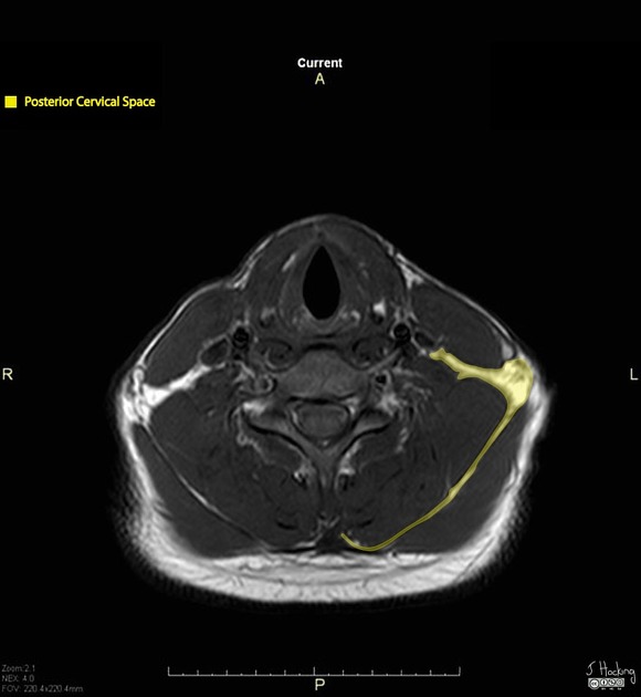
Posterior cervical space
Citation, DOI, disclosures and article data
Citation:
Lawrence N, Deng F, Knipe H, Posterior cervical space. Reference article, Radiopaedia.org (Accessed on 21 Mar 2025) https://doi.org/10.53347/rID-46968
rID:
46968
Article created:
28 Jul 2016,
Natalie Lawrence
Disclosures:
At the time the article was created Natalie Lawrence had no recorded disclosures.
View Natalie Lawrence's current disclosures
Last revised:
Disclosures:
At the time the article was last revised Francis Deng had no recorded disclosures.
View Francis Deng's current disclosures
Revisions:
6 times, by
3 contributors -
see full revision history and disclosures
Systems:
Sections:
Tags:
The posterior cervical space is one of the fat filled deep spaces of the neck located posterolaterally.
Gross Anatomy
Location
- posterolateral part of the neck extending from the mastoid tip and base of skull to the clavicles 1,2
- most of the volume is in the infrahyoid neck, with some extension into the suprahyoid neck 1
- between superficial layer of the deep cervical fascia and deep layer of deep cervical fascia 1
Contents
- fat
- spinal accessory nerve (cranial nerve XI)
- preaxillary brachial plexus 3
- lymph nodes
- level IIB
- posterolateral part of level III
- posterolateral part of level IV
- level V
Relations
- superficial: sternocleidomastoid and trapezius muscles
- deep: prevertebral space
- anterior: carotid space
A lesion in posterior cervical space typically causes anteromedial displacement of the carotid space and posteromedial displacement of the prevertebral space 2.
Related pathology
- lymphadenopathy
- branchial apparatus anomalies
References
- 1. Diagnostic Imaging: Oral and Maxillofacial. Lippincott Williams & Wilkins. ISBN:193188420X. Read it at Google Books - Find it at Amazon
- 2. Parker GD, Harnsberger HR. Radiologic evaluation of the normal and diseased posterior cervical space. AJR Am J Roentgenol. 1991;157 (1): 161-5. doi:10.2214/ajr.157.1.2048512 - Pubmed citation
- 3. Flanders AE, Rao VM, Tom BM. MRI and CT Atlas of Correlative Imaging in Otolaryngology. CRC Press. ISBN:1853170372. Read it at Google Books - Find it at Amazon
Incoming Links
Related articles: Anatomy: Head and neck
- skeleton of the head and neck
-
cranial vault
- scalp (mnemonic)
- fontanelle
-
sutures
- calvarial
- facial
- frontozygomatic suture
- frontomaxillary suture
- frontolacrimal suture
- frontonasal suture
- temporozygomatic suture
- zygomaticomaxillary suture
- parietotemporal suture (parietomastoid suture)
- occipitotemporal suture (occipitomastoid suture)
- sphenofrontal suture
- sphenozygomatic suture
- spheno-occipital suture (not a true suture)
- lacrimomaxillary suture
- nasomaxillary suture
- internasal suture
- basal/internal
- skull landmarks
- frontal bone
- temporal bone
- parietal bone
- occipital bone
- skull base (foramina)
-
facial bones
- midline single bones
- paired bilateral bones
- cervical spine
- hyoid bone
- laryngeal cartilages
-
cranial vault
- muscles of the head and neck
- muscles of the tongue (mnemonic)
- muscles of mastication
-
facial muscles
- epicranius muscle
- circumorbital and palpebral muscles
- nasal muscles
-
buccolabial muscles
- elevators, retractors and evertors of the upper lip
- levator labii superioris alaeque nasalis muscle
- levator labii superioris muscle
- zygomaticus major muscle
- zygomaticus minor muscle
- levator anguli oris muscle
- malaris muscle
- risorius muscle
- depressors, retractors and evertors of the lower lip
- depressor labii inferioris muscle
- depressor anguli oris muscle
- mentalis muscle
- compound sphincter
-
orbicularis oris muscle
- incisivus labii superioris muscle
- incisivus labii inferioris muscle
-
orbicularis oris muscle
- muscle of mastication
- modiolus
- elevators, retractors and evertors of the upper lip
- muscles of the middle ear
- orbital muscles
- muscles of the soft palate
- pharyngeal muscles
- suprahyoid muscles
- infrahyoid muscles
- intrinsic muscles of the larynx
- muscles of the neck
- platysma muscle
- longus colli muscle
- longus capitis muscle
- scalenus anterior muscle
- scalenus medius muscle
- scalenus posterior muscle
- scalenus pleuralis muscle
- sternocleidomastoid muscle
-
suboccipital muscles
- rectus capitis posterior major muscle
- rectus capitis posterior minor muscle
- obliquus capitis superior muscle
- obliquus capitis inferior muscle
- accessory muscles of the neck
- deep cervical fascia
-
deep spaces of the neck
- anterior cervical space
- buccal space
- carotid space
- danger space
- deep cervical fascia
- infratemporal fossa
- masticator space
- parapharyngeal space
- stylomandibular tunnel
- parotid space
- pharyngeal (superficial) mucosal space
- perivertebral space
- posterior cervical space
- pterygopalatine fossa
- retropharyngeal space
- suprasternal space (of Burns)
- visceral space
- surgical triangles of the neck
- orbit
- ear
- paranasal sinuses
- upper respiratory tract
- viscera of the neck
- blood supply of the head and neck
-
arterial supply
-
common carotid artery
- carotid body
- carotid bifurcation
- subclavian artery
- variants
-
common carotid artery
- venous drainage
-
arterial supply
- innervation of the head and neck
-
cranial nerves
- olfactory nerve (CN I)
- optic nerve (CN II)
- oculomotor nerve (CN III)
- trochlear nerve (CN IV)
-
trigeminal nerve (CN V) (mnemonic)
- trigeminal ganglion
- ophthalmic division
- maxillary division
- mandibular division
- abducens nerve (CN VI)
- facial nerve (CN VII)
-
vestibulocochlear nerve (CN VIII)
- vestibular ganglion (Scarpa's ganglion)
- glossopharyngeal nerve (CN IX)
- vagus nerve (CN X)
- (spinal) accessory nerve (CN XI)
- hypoglossal nerve (CN XII)
- parasympathetic ganglia of the head and neck
- cervical sympathetic ganglia
- greater occipital nerve
- third occipital nerve
-
cervical plexus
- muscular branches
- longus capitis
- longus colli
- scalenes
- geniohyoid
- thyrohyoid
-
ansa cervicalis
- omohyoid (superior and inferior bellies separately)
- sternothyroid
- sternohyoid
- phrenic nerve
- contribution to the accessory nerve (CN XI)
- cutaneous branches
- muscular branches
- brachial plexus
- pharyngeal plexus
-
cranial nerves
- lymphatic drainage of the head and neck
- embryological development of the head and neck


 Unable to process the form. Check for errors and try again.