
Pyramidal lobe of thyroid
Citation, DOI, disclosures and article data
At the time the article was created Mohammed Al Khader.O.Thabet had no recorded disclosures.
View Mohammed Al Khader.O.Thabet's current disclosuresAt the time the article was last revised Henry Knipe had the following disclosures:
- Micro-X Ltd, Shareholder (past)
These were assessed during peer review and were determined to not be relevant to the changes that were made.
View Henry Knipe's current disclosures- Pyramidal lobe of the thyroid gland
- Pyramidal lobe of the thyroid
- Pyramidal lobe of thyroid gland
- Lalouette's pyramid
- Lalouette pyramid
- Pyramidal lobe of thyroid
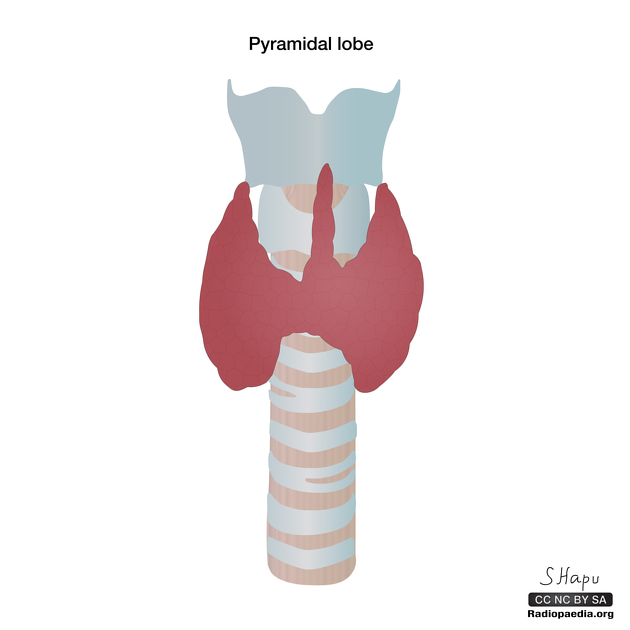
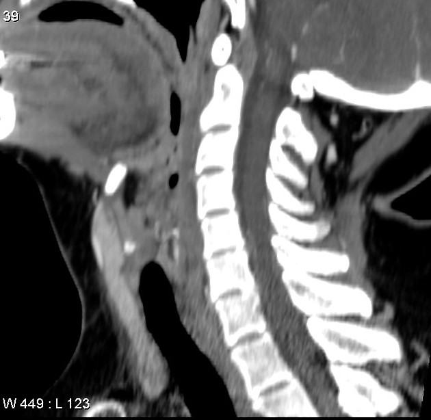
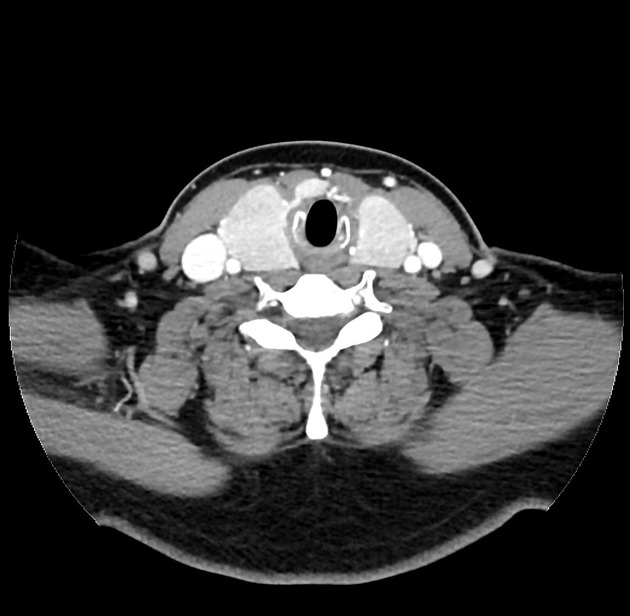
The pyramidal lobe of the thyroid, also known as Lalouette pyramid 5, is a normal anatomic variant representing a superior projection of thyroid parenchyma arising from the thyroid isthmus.
On this page:
Epidemiology
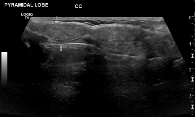
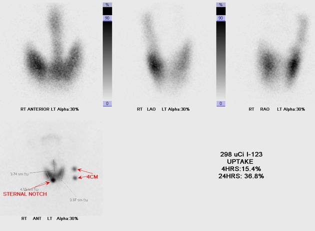
A pyramidal lobe is present in ~20 (range 10-30%) of the population ref and is commonly seen on routine thyroid ultrasound 3.
Gross anatomy
The pyramidal lobe is seen as a third thyroid lobe and represents a persistent remnant of the thyroglossal duct. It usually arises from the right or left side of the isthmus extending in a cranial direction; pyramidal lobes arising directly from the midline of the isthmus are rare (~2%) 3.
A band of fibrous tissue may be present extending superiorly from the pyramidal lobe to the hyoid bone, sometimes with a skeletal muscle component, termed the levator glandulae thyroideae muscle 4.
Clinical importance
The pathologies seen in the remainder of thyroid can also seen in the pyramidal lobe ref.
References
- 1. Anil T. Ahuja, Rhodri M. Evans. Practical Head and Neck Ultrasound. (2000) ISBN: 9781900151993 - Google Books
- 2. Wolfgang Dähnert. Radiology Review Manual. (2011) ISBN: 9781609139438 - Google Books
- 3. Mortensen C, Lockyer H, Loveday E. The Incidence and Morphological Features of Pyramidal Lobe on Thyroid Ultrasound. Ultrasound. 2014;22(4):192-8. doi:10.1177/1742271X14554677 - Pubmed
- 4. Chaudhary P, Singh Z, Khullar M, Arora K. Levator Glandulae Thyroideae, a Fibromusculoglandular Band with Absence of Pyramidal Lobe and Its Innervation: A Case Report. J Clin Diagn Res. 2013;7(7):1421-4. doi:10.7860/JCDR/2013/6144.3186 - Pubmed
- 5. Germano A, Schmitt W, Carvalho M, Marques R. Normal Ultrasound Anatomy and Common Anatomical Variants of the Thyroid Gland Plus Adjacent Structures - A Pictorial Review. Clin Imaging. 2019;58:114-28. doi:10.1016/j.clinimag.2019.07.002 - Pubmed
Incoming Links
Related articles: Anatomy: Head and neck
- skeleton of the head and neck
-
cranial vault
- scalp (mnemonic)
- fontanelle
-
sutures
- calvarial
- facial
- frontozygomatic suture
- frontomaxillary suture
- frontolacrimal suture
- frontonasal suture
- temporozygomatic suture
- zygomaticomaxillary suture
- parietotemporal suture (parietomastoid suture)
- occipitotemporal suture (occipitomastoid suture)
- sphenofrontal suture
- sphenozygomatic suture
- spheno-occipital suture (not a true suture)
- lacrimomaxillary suture
- nasomaxillary suture
- internasal suture
- basal/internal
- skull landmarks
- frontal bone
- temporal bone
- parietal bone
- occipital bone
- skull base (foramina)
-
facial bones
- midline single bones
- paired bilateral bones
- cervical spine
- hyoid bone
- laryngeal cartilages
-
cranial vault
- muscles of the head and neck
- muscles of the tongue (mnemonic)
- muscles of mastication
-
facial muscles
- epicranius muscle
- circumorbital and palpebral muscles
- nasal muscles
-
buccolabial muscles
- elevators, retractors and evertors of the upper lip
- levator labii superioris alaeque nasalis muscle
- levator labii superioris muscle
- zygomaticus major muscle
- zygomaticus minor muscle
- levator anguli oris muscle
- malaris muscle
- risorius muscle
- depressors, retractors and evertors of the lower lip
- depressor labii inferioris muscle
- depressor anguli oris muscle
- mentalis muscle
- compound sphincter
-
orbicularis oris muscle
- incisivus labii superioris muscle
- incisivus labii inferioris muscle
-
orbicularis oris muscle
- muscle of mastication
- modiolus
- elevators, retractors and evertors of the upper lip
- muscles of the middle ear
- orbital muscles
- muscles of the soft palate
- pharyngeal muscles
- suprahyoid muscles
- infrahyoid muscles
- intrinsic muscles of the larynx
- muscles of the neck
- platysma muscle
- longus colli muscle
- longus capitis muscle
- scalenus anterior muscle
- scalenus medius muscle
- scalenus posterior muscle
- scalenus pleuralis muscle
- sternocleidomastoid muscle
-
suboccipital muscles
- rectus capitis posterior major muscle
- rectus capitis posterior minor muscle
- obliquus capitis superior muscle
- obliquus capitis inferior muscle
- accessory muscles of the neck
- deep cervical fascia
-
deep spaces of the neck
- anterior cervical space
- buccal space
- carotid space
- danger space
- deep cervical fascia
- infratemporal fossa
- masticator space
- parapharyngeal space
- stylomandibular tunnel
- parotid space
- pharyngeal (superficial) mucosal space
- perivertebral space
- posterior cervical space
- pterygopalatine fossa
- retropharyngeal space
- suprasternal space (of Burns)
- visceral space
- surgical triangles of the neck
- orbit
- ear
- paranasal sinuses
- upper respiratory tract
- viscera of the neck
- blood supply of the head and neck
-
arterial supply
-
common carotid artery
- carotid body
- carotid bifurcation
- subclavian artery
- variants
-
common carotid artery
- venous drainage
-
arterial supply
- innervation of the head and neck
-
cranial nerves
- olfactory nerve (CN I)
- optic nerve (CN II)
- oculomotor nerve (CN III)
- trochlear nerve (CN IV)
-
trigeminal nerve (CN V) (mnemonic)
- trigeminal ganglion
- ophthalmic division
- maxillary division
- mandibular division
- abducens nerve (CN VI)
- facial nerve (CN VII)
-
vestibulocochlear nerve (CN VIII)
- vestibular ganglion (Scarpa's ganglion)
- glossopharyngeal nerve (CN IX)
- vagus nerve (CN X)
- (spinal) accessory nerve (CN XI)
- hypoglossal nerve (CN XII)
- parasympathetic ganglia of the head and neck
- cervical sympathetic ganglia
- greater occipital nerve
- third occipital nerve
-
cervical plexus
- muscular branches
- longus capitis
- longus colli
- scalenes
- geniohyoid
- thyrohyoid
-
ansa cervicalis
- omohyoid (superior and inferior bellies separately)
- sternothyroid
- sternohyoid
- phrenic nerve
- contribution to the accessory nerve (CN XI)
- cutaneous branches
- muscular branches
- brachial plexus
- pharyngeal plexus
-
cranial nerves
- lymphatic drainage of the head and neck
- embryological development of the head and neck


 Unable to process the form. Check for errors and try again.