
Retropubic space
Citation, DOI, disclosures and article data
At the time the article was created Ayush Goel had no recorded disclosures.
View Ayush Goel's current disclosuresAt the time the article was last revised Mohd Ashyiraff Ilani Bin Ismail had no financial relationships to ineligible companies to disclose.
View Mohd Ashyiraff Ilani Bin Ismail's current disclosures- Cave of Retzius
- Prevesical space
- Cavum Retzii
- Space of Retzius
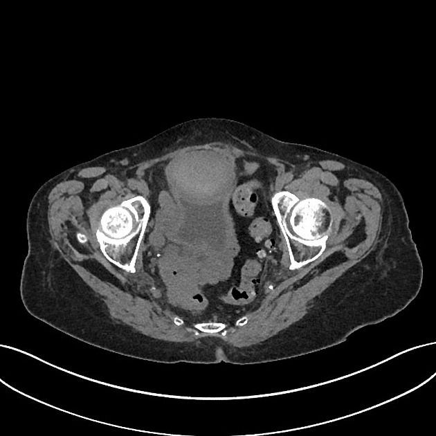
The retropubic space (also known as the prevesical space, cave of Retzius or cavum Retzii) is an extraperitoneal space located posterior to the pubic symphysis and anterior to the urinary bladder. It is separated from the anterior abdominal wall by the transversalis fascia and extends to the level of the umbilicus.
It is an avascular potential space used as prosthesis reservoir for penile implant procedure and also used for stress urinary incontinence procedure in urogynecology5.
History and etymology
This space is named after Anders Retzius (1796–1860), a Swedish professor of anatomy 4.
Related pathology
References
- 1. Sectional Anatomy for Imaging Professionals. Mosby. ISBN:0323082602. Read it at Google Books - Find it at Amazon
- 2. Butler P, Mitchell A, Healy JC. Applied Radiological Anatomy. Cambridge University Press. (2012) ISBN:0521766664. Read it at Google Books - Find it at Amazon
- 3. O'Connell AM, Duddy L, Lee C, Lee MJ. CT of pelvic extraperitoneal spaces: an anatomical study in cadavers. Clinical radiology. 62 (5): 432-8. doi:10.1016/j.crad.2006.11.012 - Pubmed
- 4. Anders Retzius (1796–1860). (2013) Journal of Neurology. 260 (5): 1445. doi:10.1007/s00415-012-6728-7 - Pubmed
- 5. Patel J, Sasson A, Simpson W, Wilck E. The Anatomy and Pathology of the Space of Retzius. Clin Imaging. 2024;110:110137. doi:10.1016/j.clinimag.2024.110137 - Pubmed
Incoming Links
Related articles: Anatomy: Abdominopelvic
- skeleton of the abdomen and pelvis
- muscles of the abdomen and pelvis
- spaces of the abdomen and pelvis
- anterior abdominal wall
- posterior abdominal wall
- abdominal cavity
- pelvic cavity
- perineum
- abdominal and pelvic viscera
- gastrointestinal tract
- spleen
- hepatobiliary system
-
endocrine system
-
adrenal gland
- adrenal vessels
- chromaffin cells
- variants
- pancreas
- organs of Zuckerkandl
-
adrenal gland
-
urinary system
-
kidney
- renal pelvis
- renal sinus
- avascular plane of Brodel
-
variants
- number
- fusion
- location
- shape
- ureter
- urinary bladder
- urethra
- embryology
-
kidney
- male reproductive system
-
female reproductive system
- vulva
- vagina
- uterus
- adnexa
- Fallopian tubes
- ovaries
- broad ligament (mnemonic)
- variant anatomy
- embryology
- blood supply of the abdomen and pelvis
- arteries
-
abdominal aorta
- inferior phrenic artery
- celiac artery
- superior mesenteric artery
- middle suprarenal artery
- renal artery (variant anatomy)
- gonadal artery (ovarian artery | testicular artery)
- inferior mesenteric artery
- lumbar arteries
- median sacral artery
-
common iliac artery
- external iliac artery
-
internal iliac artery (mnemonic)
- anterior division
- umbilical artery
- superior vesical artery
- obturator artery
- vaginal artery
- inferior vesical artery
- uterine artery
- middle rectal artery
-
internal pudendal artery
- inferior rectal artery
-
perineal artery
- posterior scrotal artery
- transverse perineal artery
- artery to the bulb
- deep artery of the penis/clitoris
- dorsal artery of the penis/clitoris
- inferior gluteal artery
- posterior division (mnemonic)
- variant anatomy
- anterior division
-
abdominal aorta
- portal venous system
- veins
- anastomoses
- arterioarterial anastomoses
- portal-systemic venous collateral pathways
- watershed areas
- arteries
- lymphatics
- innervation of the abdomen and pelvis
- thoracic splanchnic nerves
- lumbar plexus
-
sacral plexus
- lumbosacral trunk
- sciatic nerve
- superior gluteal nerve
- inferior gluteal nerve
- nerve to piriformis
- perforating cutaneous nerve
- posterior femoral cutaneous nerve
- parasympathetic pelvic splanchnic nerves
- pudendal nerve
- nerve to quadratus femoris and inferior gemellus muscles
- nerve to internal obturator and superior gemellus muscles
- autonomic ganglia and plexuses


 Unable to process the form. Check for errors and try again.