
Temporomandibular joint disc
Citation, DOI, disclosures and article data
At the time the article was created Frank Gaillard had no recorded disclosures.
View Frank Gaillard's current disclosuresAt the time the article was last revised Craig Hacking had the following disclosures:
- Philips Australia, Paid speaker at Philips Spectral CT events (ongoing)
These were assessed during peer review and were determined to not be relevant to the changes that were made.
View Craig Hacking's current disclosures- TMJ disc
The temporomandibular joint (TMJ) disc (or meniscus) is made of fibrocartilage and divides the temporomandibular joint into two compartments.
Gross anatomy
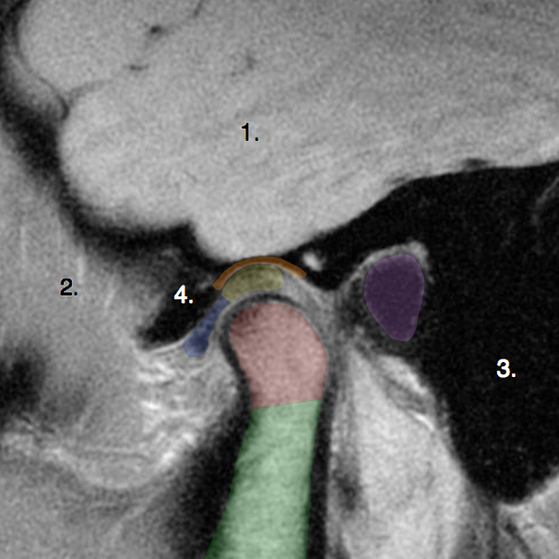
The disc is composed of fibrocartilage, with crimped collagen, thought to better absorb impact. It has a biconcave shape with a thicker periphery and attached at its periphery (except posteriorly) to the TMJ capsule (see below). There are anterior and posterior transverse thickenings of the disc, known as the anterior and posterior bands. Between the two is the intermediate zone.
The TMJ disc divides the joint into two compartments which separates translational and rotational motion:
superior discotemporal space: located above the disc, between it and the mandibular fossa and articular eminence of the temporal bone. Anterior translation during mouth opening occurs here.
inferior discomandibular space: located below the disc, between it and the mandibular condyle. Rotation occurs here (in absence of TMJ dysfunction).
Attachments
anterior: capsule which blends with the insertion of the superior belly lateral pterygoid muscle, and attaches to the articular eminence of the temporal bone superiorly and neck of condyle inferiorly
medial: capsule
lateral: capsule
posterior: blends with the retrodiscal layer
Radiographic features
MRI
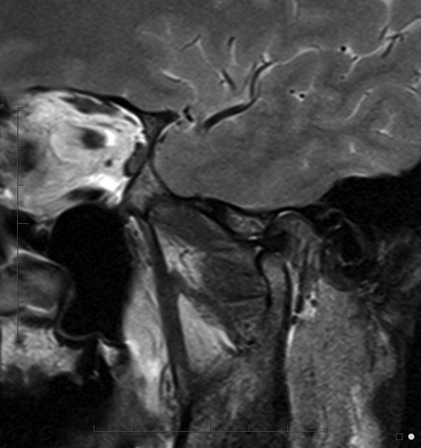
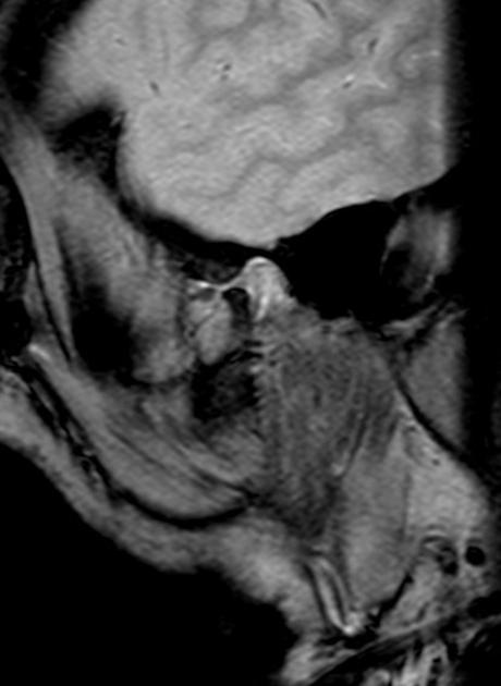
The anterior band and intermediate zone are low signal on T2/PD sequences, whereas the posterior band and retrodiscal zone are intermediate to hyperintense. The intermediate zone may have a higher T2 signal cleft within it as a normal variant.
The anterior band is well seen on both open and closed mouth oblique sagittal views (see: TMJ MRI protocol), whereas the posterior band and retrodiscal zone are best seen in closed mouth views.
A normal disc is biconcave. In pathology this morphology alters, and the disc may appear
thickened and globular / lentiform
irregular
thinned
perforated
References
- 1. Oliver J. Sommer, Felix Aigner, Ansgar Rudisch, Hannes Gruber, Helga Fritsch, Werner Millesi, and Michael Stiskal "Cross-sectional and Functional Imaging of the Temporomandibular Joint: Radiology, Pathology, and Basic Biomechanics of the Jaw" RadioGraphics 2003 23: e14; published online as 10.1148/rg.e14
- 2. Xavier Tomas, Jaume Pomes, Juan Berenguer, Llorenç Quinto, Carlos Nicolau, Josep Maria Mercader, and Vicente Castro "MR Imaging of Temporomandibular Joint Dysfunction: A Pictorial Review" RadioGraphics 2006 26: 765-781.
- 3. Standring S (editor). Gray's Anatomy (39th edition). Churchill Livingstone. (2011) ISBN:0443066841. Read it at Google Books - Find it at Amazon
Incoming Links
- Temporomandibular joint dysfunction - anterior disc displacement with reduction
- Temporomandibular joint dysfunction
- Temporomandibular joints anterior disc displacement with reduction
- Apparent discontinuity of the roof of the mandibular fossa
- Temporomandibular joint with anterior disc dislocation and recapture
- Irreducible anterior temporomandibular disc dislocation - bilateral
- Temporomandibular joint with dislocated disc and recapture
- Temporomandibular joint with dislocated disc and recapture
- Unilateral temporomandibular disc displacement without recapture
- Normal temporomandibular joint
- Anterior temporomandibular joint disk displacement with recapture
- Hypertranslation of mandibular condyle
- Irreducible anterior dislocation - TMJ disc
- Irreducible anterior temporomandibular disc dislocation - bilateral
- Temporomandibular joint (annotated MRI)
- TMJ - retrodiscal area (annotated MRI)
- Junction of intermediate zone and posterior band
- Temporomandibular joint - stuck disc
- Irreducible anterior dislocation - TMJ disc
- Temporomandibular joint: sclerosed mandibular condyle and ill-defined disc
Related articles: Anatomy: Head and neck
- skeleton of the head and neck
-
cranial vault
- scalp (mnemonic)
- fontanelle
-
sutures
- calvarial
- facial
- frontozygomatic suture
- frontomaxillary suture
- frontolacrimal suture
- frontonasal suture
- temporozygomatic suture
- zygomaticomaxillary suture
- parietotemporal suture (parietomastoid suture)
- occipitotemporal suture (occipitomastoid suture)
- sphenofrontal suture
- sphenozygomatic suture
- spheno-occipital suture (not a true suture)
- lacrimomaxillary suture
- nasomaxillary suture
- internasal suture
- basal/internal
- skull landmarks
- frontal bone
- temporal bone
- parietal bone
- occipital bone
- skull base (foramina)
-
facial bones
- midline single bones
- paired bilateral bones
- cervical spine
- hyoid bone
- laryngeal cartilages
-
cranial vault
- muscles of the head and neck
- muscles of the tongue (mnemonic)
- muscles of mastication
-
facial muscles
- epicranius muscle
- circumorbital and palpebral muscles
- nasal muscles
-
buccolabial muscles
- elevators, retractors and evertors of the upper lip
- levator labii superioris alaeque nasalis muscle
- levator labii superioris muscle
- zygomaticus major muscle
- zygomaticus minor muscle
- levator anguli oris muscle
- malaris muscle
- risorius muscle
- depressors, retractors and evertors of the lower lip
- depressor labii inferioris muscle
- depressor anguli oris muscle
- mentalis muscle
- compound sphincter
-
orbicularis oris muscle
- incisivus labii superioris muscle
- incisivus labii inferioris muscle
-
orbicularis oris muscle
- muscle of mastication
- modiolus
- elevators, retractors and evertors of the upper lip
- muscles of the middle ear
- orbital muscles
- muscles of the soft palate
- pharyngeal muscles
- suprahyoid muscles
- infrahyoid muscles
- intrinsic muscles of the larynx
- muscles of the neck
- platysma muscle
- longus colli muscle
- longus capitis muscle
- scalenus anterior muscle
- scalenus medius muscle
- scalenus posterior muscle
- scalenus pleuralis muscle
- sternocleidomastoid muscle
-
suboccipital muscles
- rectus capitis posterior major muscle
- rectus capitis posterior minor muscle
- obliquus capitis superior muscle
- obliquus capitis inferior muscle
- accessory muscles of the neck
- deep cervical fascia
-
deep spaces of the neck
- anterior cervical space
- buccal space
- carotid space
- danger space
- deep cervical fascia
- infratemporal fossa
- masticator space
- parapharyngeal space
- stylomandibular tunnel
- parotid space
- pharyngeal (superficial) mucosal space
- perivertebral space
- posterior cervical space
- pterygopalatine fossa
- retropharyngeal space
- suprasternal space (of Burns)
- visceral space
- surgical triangles of the neck
- orbit
- ear
- paranasal sinuses
- upper respiratory tract
- viscera of the neck
- blood supply of the head and neck
-
arterial supply
-
common carotid artery
- carotid body
- carotid bifurcation
- subclavian artery
- variants
-
common carotid artery
- venous drainage
-
arterial supply
- innervation of the head and neck
-
cranial nerves
- olfactory nerve (CN I)
- optic nerve (CN II)
- oculomotor nerve (CN III)
- trochlear nerve (CN IV)
-
trigeminal nerve (CN V) (mnemonic)
- trigeminal ganglion
- ophthalmic division
- maxillary division
- mandibular division
- abducens nerve (CN VI)
- facial nerve (CN VII)
-
vestibulocochlear nerve (CN VIII)
- vestibular ganglion (Scarpa's ganglion)
- glossopharyngeal nerve (CN IX)
- vagus nerve (CN X)
- (spinal) accessory nerve (CN XI)
- hypoglossal nerve (CN XII)
- parasympathetic ganglia of the head and neck
- cervical sympathetic ganglia
- greater occipital nerve
- third occipital nerve
-
cervical plexus
- muscular branches
- longus capitis
- longus colli
- scalenes
- geniohyoid
- thyrohyoid
-
ansa cervicalis
- omohyoid (superior and inferior bellies separately)
- sternothyroid
- sternohyoid
- phrenic nerve
- contribution to the accessory nerve (CN XI)
- cutaneous branches
- muscular branches
- brachial plexus
- pharyngeal plexus
-
cranial nerves
- lymphatic drainage of the head and neck
- embryological development of the head and neck


 Unable to process the form. Check for errors and try again.