
Buccal space
Citation, DOI, disclosures and article data
At the time the article was created Craig Hacking had no recorded disclosures.
View Craig Hacking's current disclosuresAt the time the article was last revised Tariq Walizai had no financial relationships to ineligible companies to disclose.
View Tariq Walizai's current disclosures- Buccinator space
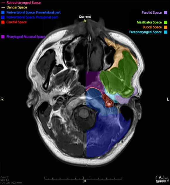
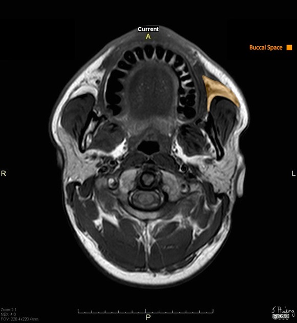
The buccal space, also known as the buccinator space, is one of the seven suprahyoid deep compartments of the head and neck.
Gross anatomy
The buccal spaces are paired fat-containing spaces on each side of the face forming the cheeks. Each space is enveloped by the superficial (investing) layer of the deep cervical fascia.
It is located between the buccinator and platysma muscles, therefore it is only a small potential space with limited contents.
Contents
fat: cheek padding
accessory parotid gland in 20% of people which can cause facial asymmetry; readily seen on MRI
facial and buccal arteries and corresponding veins
facial nerve (CN VII): buccal branch
trigeminal nerve (CN V): buccal nerve of the mandibular division
Boundaries and relations
anterior: orbicularis oris muscle and the angle of the mouth
posterior: masseter muscle, mandible, medial pterygoid and lateral pterygoid muscles
superior: zygomatic process of the maxilla and zygomaticus muscles
inferior: depressor anguli oris muscle and the deep fascia attaching to the mandible
medial (deep): buccinator muscle
lateral (superficial): platysma muscle and subcutaneous tissues with the skin
Communications
Buccal space infection can spread to/from the teeth. There is no real boundary between the buccal space and the submandibular space inferiorly. There is also potential communication with the pterygomandibular region, infratemporal space, and the parapharyngeal space posteriorly.
Related pathology
tumors: minor salivary gland tumors, vascular lesions (e.g. hemangiomas)
References
- 1. Harnsberger HR, Glastonbury CM, Michel MA et-al. Diagnostic Imaging: Head and Neck. Lippincott Williams & Wilkins. (2010) ISBN:1931884781. Read it at Google Books - Find it at Amazon
- 2. Last's anatomy, regional and applied. Churchill Livingstone. ISBN:044304662X. Read it at Google Books - Find it at Amazon
- 3. Moore KL, Agur AMR, Dalley AF. Clinically oriented anatomy. LWW. ISBN:1451119453. Read it at Google Books - Find it at Amazon
- 4. Tart RP, Kotzur IM, Mancuso AA et-al. CT and MR imaging of the buccal space and buccal space masses. Radiographics. 1995;15 (3): 531-50. doi:10.1148/radiographics.15.3.7624561 - Pubmed citation
Incoming Links
- Buccal mucosal squamous cell carcinoma
- Adenoid cystic carcinoma of maxillary sinus
- Chondrosarcoma of the maxilla
- Buccal squamous cell carcinoma
- Sinonasal lymphoma
- Haemangioma - buccal space
- Buccal fat pad arteriovenous malformation
- Maxillary sinus squamous cell carcinoma
- Buccal space (annotated MRI)
- Buccal squamous cell carcinoma
- Buccal squamous cell carcinoma
Related articles: Anatomy: Head and neck
- skeleton of the head and neck
-
cranial vault
- scalp (mnemonic)
- fontanelle
-
sutures
- calvarial
- facial
- frontozygomatic suture
- frontomaxillary suture
- frontolacrimal suture
- frontonasal suture
- temporozygomatic suture
- zygomaticomaxillary suture
- parietotemporal suture (parietomastoid suture)
- occipitotemporal suture (occipitomastoid suture)
- sphenofrontal suture
- sphenozygomatic suture
- spheno-occipital suture (not a true suture)
- lacrimomaxillary suture
- nasomaxillary suture
- internasal suture
- basal/internal
- skull landmarks
- frontal bone
- temporal bone
- parietal bone
- occipital bone
- skull base (foramina)
-
facial bones
- midline single bones
- paired bilateral bones
- cervical spine
- hyoid bone
- laryngeal cartilages
-
cranial vault
- muscles of the head and neck
- muscles of the tongue (mnemonic)
- muscles of mastication
-
facial muscles
- epicranius muscle
- circumorbital and palpebral muscles
- nasal muscles
-
buccolabial muscles
- elevators, retractors and evertors of the upper lip
- levator labii superioris alaeque nasalis muscle
- levator labii superioris muscle
- zygomaticus major muscle
- zygomaticus minor muscle
- levator anguli oris muscle
- malaris muscle
- risorius muscle
- depressors, retractors and evertors of the lower lip
- depressor labii inferioris muscle
- depressor anguli oris muscle
- mentalis muscle
- compound sphincter
-
orbicularis oris muscle
- incisivus labii superioris muscle
- incisivus labii inferioris muscle
-
orbicularis oris muscle
- muscle of mastication
- modiolus
- elevators, retractors and evertors of the upper lip
- muscles of the middle ear
- orbital muscles
- muscles of the soft palate
- pharyngeal muscles
- suprahyoid muscles
- infrahyoid muscles
- intrinsic muscles of the larynx
- muscles of the neck
- platysma muscle
- longus colli muscle
- longus capitis muscle
- scalenus anterior muscle
- scalenus medius muscle
- scalenus posterior muscle
- scalenus pleuralis muscle
- sternocleidomastoid muscle
-
suboccipital muscles
- rectus capitis posterior major muscle
- rectus capitis posterior minor muscle
- obliquus capitis superior muscle
- obliquus capitis inferior muscle
- accessory muscles of the neck
- deep cervical fascia
-
deep spaces of the neck
- anterior cervical space
- buccal space
- carotid space
- danger space
- deep cervical fascia
- infratemporal fossa
- masticator space
- parapharyngeal space
- stylomandibular tunnel
- parotid space
- pharyngeal (superficial) mucosal space
- perivertebral space
- posterior cervical space
- pterygopalatine fossa
- retropharyngeal space
- suprasternal space (of Burns)
- visceral space
- surgical triangles of the neck
- orbit
- ear
- paranasal sinuses
- upper respiratory tract
- viscera of the neck
- blood supply of the head and neck
-
arterial supply
-
common carotid artery
- carotid body
- carotid bifurcation
- subclavian artery
- variants
-
common carotid artery
- venous drainage
-
arterial supply
- innervation of the head and neck
-
cranial nerves
- olfactory nerve (CN I)
- optic nerve (CN II)
- oculomotor nerve (CN III)
- trochlear nerve (CN IV)
-
trigeminal nerve (CN V) (mnemonic)
- trigeminal ganglion
- ophthalmic division
- maxillary division
- mandibular division
- abducens nerve (CN VI)
- facial nerve (CN VII)
-
vestibulocochlear nerve (CN VIII)
- vestibular ganglion (Scarpa's ganglion)
- glossopharyngeal nerve (CN IX)
- vagus nerve (CN X)
- (spinal) accessory nerve (CN XI)
- hypoglossal nerve (CN XII)
- parasympathetic ganglia of the head and neck
- cervical sympathetic ganglia
- greater occipital nerve
- third occipital nerve
-
cervical plexus
- muscular branches
- longus capitis
- longus colli
- scalenes
- geniohyoid
- thyrohyoid
-
ansa cervicalis
- omohyoid (superior and inferior bellies separately)
- sternothyroid
- sternohyoid
- phrenic nerve
- contribution to the accessory nerve (CN XI)
- cutaneous branches
- muscular branches
- brachial plexus
- pharyngeal plexus
-
cranial nerves
- lymphatic drainage of the head and neck
- embryological development of the head and neck


 Unable to process the form. Check for errors and try again.