
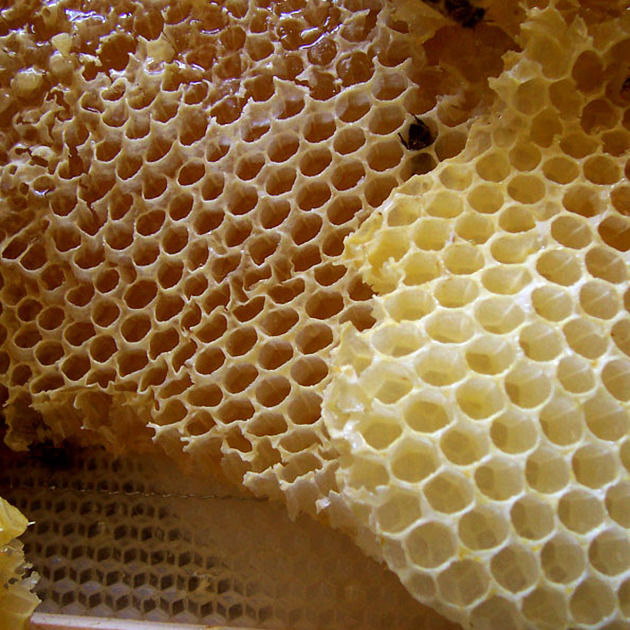
Honeycombing describes small adjacent subpleural cystic structures in the context of established pulmonary fibrosis with destruction of lung parenchyma and loss of architecture 11. A single layer of cysts is now considered sufficient to apply this descriptor if other signs of pulmonary fibrosis are present 11.
On this page:
Terminology
The terms microcystic honeycombing and macrocystic honeycombing are sometimes used but without a clear definition. The initial stages of honeycombing are perceptible only on microscopy. Cysts progressively enlarge, becoming visible on CT 11. Exuberant honeycombing refers to numerous large adjacent cysts and is a feature of connective tissue disease 13.
Pathology
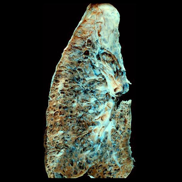
The typical histological correlate of honeycombing is advanced traction bronchiolectasis surrounded by alveolar collapse 9. Pathologists regard honeycomb cysts as collapsed cystic secondary lobules lined by respiratory epithelium 12. Cysts are typically connected to airways by a slit-like orifice and flap which suggests the role of a check- valve mechanism in cyst growth. Rapid pre-terminal cyst growth has been reported with autopsy correlation 14.
Radiographic features
CT
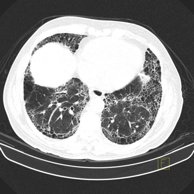
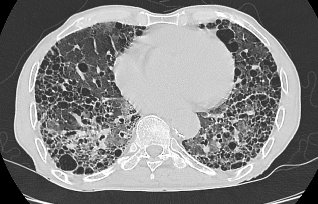
Honeycombing and traction bronchiolectasis can be difficult to distinguish, but this is not surprising since traction bronchiolectasis is thought to progress to honeycombing. Honeycomb cysts are usually similar in size, <5 mm (range 3-25 mm), clustered in a row or stacked and with shared walls. The walls are relatively thick, around 1-3 mm. They are first seen adjacent to the pleura, usually in the basal lower lobes in areas of dense fibrosis 10. The cysts enlarge over time and acute pre-terminal enlargement has been described in one patient with rapid deccline in pulmonary function and autopsy features supporting a check-valve mechanism
History and etymology
The term “honeycomb lung” is thought to have originated in the 19th century in Germany and is thought to have first appeared in 1949 in a study by N Oswald and T Parkinson 5.
Differential diagnosis
-
in some situations
paraseptal emphysema (single layer of larger thin-walled juxta-pleural cyst-like spaces in non-fibrotic lung with upper lobe predominance) 10
airspace enlargement with fibrosis/smoking-related interstitial fibrosis (larger multi-layered or confluent thin-walled cysts, or cysts separated by normal lung which often spare the subpleural lung and lower zones) 10
airspace consolidation in the presence of pulmonary emphysema 8
Practical points
using the term "honeycombing" implies substantial pulmonary fibrosis is present, so it should be used with care 11
differentiation of honeycombing from emphysematous spaces with peripheral fibrosis can be difficult and the two conditions may coexist; the distribution is helpful, honeycombing is posterior lower zone predominant whereas emphysema with fibrosis is upper zone predominant 12
pathologists may find the diagnosis of honeycombing difficult as it can be difficult to distinguish from traction bronchiectasis; a clear histological definition would facilitate imaging evaluation 12


 Unable to process the form. Check for errors and try again.