
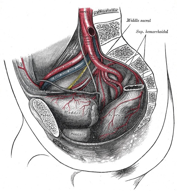
The internal iliac artery (also known as the hypogastric artery, but internal iliac is the accepted term in the TA) is the smaller terminal branch of the common iliac artery. It supplies the pelvic walls, pelvic viscera, external genitalia, perineum, buttock and medial part of the thigh.
On this page:
Gross anatomy
Origin
The common iliac artery bifurcates into the internal iliac artery and external iliac artery at the level of the pelvic brim anterior to the sacroiliac joint.
Course
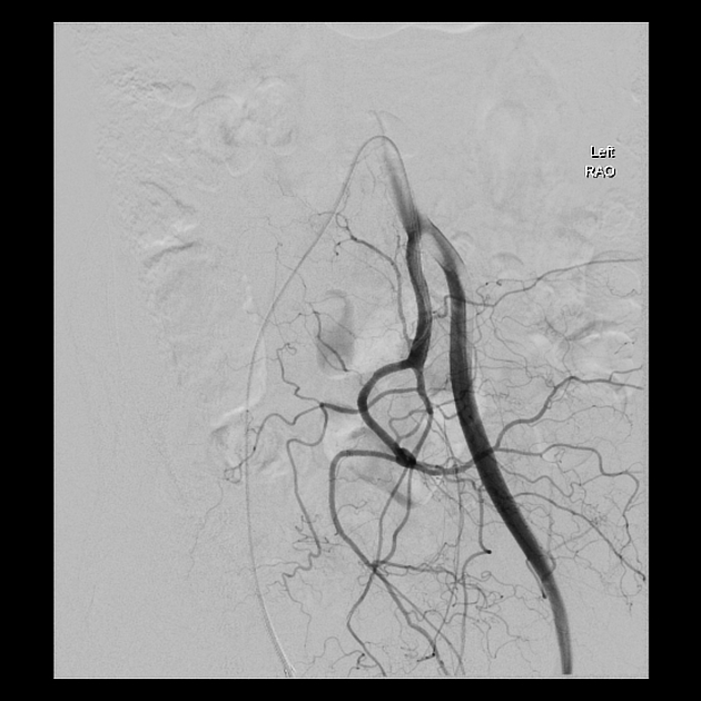
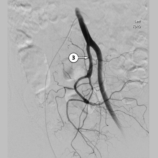
The internal iliac artery courses posteromedially towards the greater sciatic foramen. It is approximately 4 cm in length.
At the superior margin of the greater sciatic foramen it divides into an anterior division and posterior division. The anterior division continues down to the ischial spine anterior to piriformis giving off visceral and parietal branches. The posterior division only gives rise to parietal branches.
Branches
Anterior division
- umbilical artery (only patent in the fetus)
- superior vesical artery (branch of the umbilical artery)
- obturator artery (25% will branch off the inferior epigastric artery)
- vaginal artery
- inferior vesical artery
- uterine artery
- middle rectal artery
- internal pudendal artery (supplies the external genitalia)
- inferior gluteal artery
The obturator, internal pudendal and inferior gluteal arteries are parietal branches, whereas the other arteries in the above list are visceral arteries (i.e. umbilical, superior and inferior vesical, vaginal, uterine and middle rectal artery).
The nine branches of the anterior division of the internal iliac artery may be more easily remembered in these divisions:
- "three urinary": umbilical artery, superior vesical artery, inferior vesical artery
- "three visceral": uterine artery, vaginal artery, middle rectal artery
- "three parietal": obturator artery, internal pudendal artery, inferior gluteal artery
Posterior division
- iliolumbar artery
-
lateral sacral arteries
- superior and inferior branches
- superior gluteal artery
Mnemonics to remember branches of internal iliac artery include:
- I Love Sex or PILLS G (posterior division)
- SOI VU MR PIG (anterior division)
- I Love Going Places In My Very Own Underwear! (anterior and posterior divisions)
- I <3 U SUMO SIL (anterior and posterior divisions)
Relations
- anteriorly: ureter, ovary, uterine tube
- posteriorly: internal iliac vein, lumbosacral trunk, sacroiliac joint
- medially: peritoneum
- laterally: external iliac vein, obturator nerve 3
Variant anatomy
Radiographic features
CT
Contrast-enhanced CT is able to accurately detect many of the parietal branches of the internal iliac artery 4:
- iliolumbar artery
- courses superolaterally from the SI joint and divides into iliac and lumbar branches superior to the pelvic brim behind the psoas muscle
- lateral sacral arteries
- descend anterior to the sacrum and branches enter sacral foramina
- superior gluteal artery
- exits the pelvis via the greater sciatic foramen and here it is found posterior to the ilium and superior to piriformis muscle
- inferior gluteal artery
- posterior to the ischial spine, inferior to piriformis muscle; after exiting the pelvis it is covered by gluteus maximus muscle
- internal pudendal artery
- found posterior to the ischial spine and runs along the medial aspect of the obturator internus muscle
- obturator artery
- seen dividing into anterior and posterior branches in the obturator canal
The only visceral branches that are easily identified on CT imaging are the 4:
- uterine artery
- has a typical 'corkscrew' appearance in the broad ligament
- superior vesical artery
- found lateral to the bladder


 Unable to process the form. Check for errors and try again.