Junctional parenchymal defect of kidney
Citation, DOI, disclosures and article data
At the time the article was created Praveen Jha had no recorded disclosures.
View Praveen Jha's current disclosuresAt the time the article was last revised Mateusz Wilczek had no financial relationships to ineligible companies to disclose.
View Mateusz Wilczek's current disclosures- Renal junctional parenchymal defect
- Renal junctional parenchymal defects
- Junctional parenchymal defect of kidneys
- Junctional parenchymal defect of the kidney
- Junctional cleft of kidney
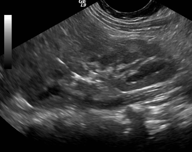
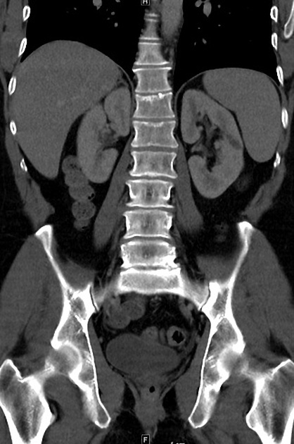
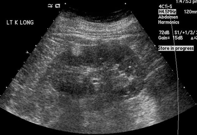
Junctional parenchymal defects in renal imaging are a normal variant, which results from the incomplete embryonic fusion of renunculi.
Radiographic features
Ultrasound
It can be seen as a triangular echogenic cortical defect, frequently seen in upper lobe parenchyma. The defect is the extension of sinus fat into the cortex, usually at the border of the upper pole and interpolar region of the kidney.
Differential diagnosis
General imaging considerations include:
renal angiomyolipoma (usually more round)
persistent fetal lobulation of the kidneys (also resulting from incomplete fusion of renal lobules)
References
- 1. Carter AR, Horgan JG, Jennings TA et-al. The junctional parenchymal defect: a sonographic variant of renal anatomy. Radiology. 1985;154 (2): 499-502. Radiology (link) - Pubmed citation
- 2. Hélénon O, Merran S, Paraf F et-al. Unusual fat-containing tumors of the kidney: a diagnostic dilemma. Radiographics. 1997;17 (1): 129-44. doi:10.1148/radiographics.17.1.9017804 - Pubmed citation
Incoming Links
Related articles: Anatomy: Abdominopelvic
- skeleton of the abdomen and pelvis
- muscles of the abdomen and pelvis
- spaces of the abdomen and pelvis
- anterior abdominal wall
- posterior abdominal wall
- abdominal cavity
- pelvic cavity
- perineum
- abdominal and pelvic viscera
- gastrointestinal tract
- spleen
- hepatobiliary system
-
endocrine system
-
adrenal gland
- adrenal vessels
- chromaffin cells
- variants
- pancreas
- organs of Zuckerkandl
-
adrenal gland
-
urinary system
-
kidney
- renal pelvis
- renal sinus
- avascular plane of Brodel
-
variants
- number
- fusion
- location
- shape
- ureter
- urinary bladder
- urethra
- embryology
-
kidney
- male reproductive system
-
female reproductive system
- vulva
- vagina
- uterus
- adnexa
- Fallopian tubes
- ovaries
- broad ligament (mnemonic)
- variant anatomy
- embryology
- blood supply of the abdomen and pelvis
- arteries
-
abdominal aorta
- inferior phrenic artery
- celiac artery
- superior mesenteric artery
- middle suprarenal artery
- renal artery (variant anatomy)
- gonadal artery (ovarian artery | testicular artery)
- inferior mesenteric artery
- lumbar arteries
- median sacral artery
-
common iliac artery
- external iliac artery
-
internal iliac artery (mnemonic)
- anterior division
- umbilical artery
- superior vesical artery
- obturator artery
- vaginal artery
- inferior vesical artery
- uterine artery
- middle rectal artery
-
internal pudendal artery
- inferior rectal artery
-
perineal artery
- posterior scrotal artery
- transverse perineal artery
- artery to the bulb
- deep artery of the penis/clitoris
- dorsal artery of the penis/clitoris
- inferior gluteal artery
- posterior division (mnemonic)
- variant anatomy
- anterior division
-
abdominal aorta
- portal venous system
- veins
- anastomoses
- arterioarterial anastomoses
- portal-systemic venous collateral pathways
- watershed areas
- arteries
- lymphatics
- innervation of the abdomen and pelvis
- thoracic splanchnic nerves
- lumbar plexus
-
sacral plexus
- lumbosacral trunk
- sciatic nerve
- superior gluteal nerve
- inferior gluteal nerve
- nerve to piriformis
- perforating cutaneous nerve
- posterior femoral cutaneous nerve
- parasympathetic pelvic splanchnic nerves
- pudendal nerve
- nerve to quadratus femoris and inferior gemellus muscles
- nerve to internal obturator and superior gemellus muscles
- autonomic ganglia and plexuses


 Unable to process the form. Check for errors and try again.