
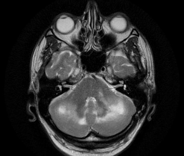
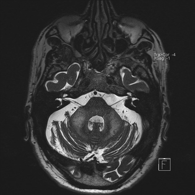
The shrimp sign is an MRI marker of cerebellar progressive multifocal leukoencephalopathy, characterized by T2-hyperintensity in the cerebellar white matter abutting but sparing the dentate nucleus.
The white matter lesion resembles a shrimp, with the dentate nucleus outlining the belly of the shrimp, the broader area of involvement at the middle cerebellar peduncle corresponding to the head end of the shrimp, and the narrower area of involvement in the medial cerebellar white matter corresponding to the tail. There are detailed diagnostic criteria to maintain reliability and accuracy of the sign 1.
On this page:
Inclusion criteria
The presence of the following features qualifies a white matter lesion as the shrimp sign 1:
well-defined
located in the cerebellar white matter
hyperintense on T2-weighted and T2-FLAIR sequences
hypointense on T1-weighted sequences
abutting and sharply outlining the dentate nucleus (in axial, sagittal, and/or coronal planes)
encompassing at least 50% of the dentate nucleus (not necessarily contiguous)
Exclusion criteria
The white matter lesion does not meet the shrimp sign if any of these features are present 1:
not hypointense on T1-weighted imaging
hazy and ill-defined on T2-weighted and T2-FLAIR sequences
cavitation within the lesion
prominent focal, diffuse, or ring enhancement
involvement of the dentate nucleus
displacement of the dentate nucleus
enhancement of the dentate nucleus
severe atrophy of the dentate nucleus early in the disease course
Optional features
Some features are compatible or permissible with the diagnosis 1:
mottled appearance of the lesion on T2-weighted imaging
involvement of the white matter hilum of the dentate nucleus
co-occurrence with or independence of cerebral and brainstem lesions
minimal enlargement (<2–3 mm) of the middle cerebellar peduncle
minimal mass effect on the fourth ventricle
faint enhancement of the white matter lesion or the hilum of the dentate nucleus
olivopontocerebellar atrophy in late-stage disease
bilateral, but usually asymmetric, white matter lesions
Differential diagnosis
Fragile X-associated tremor/ataxia syndrome, the cause of the middle cerebellar peduncle sign, can look like the shrimp sign but is typically bilateral symmetric, rather than unilateral or asymmetric like the shrimp sign. Moreover, the clinical context should help distinguish these entities whereby fragile X-associated tremor/ataxia syndrome occurs in older men with a familial history, while most cases of progressive multifocal leukoencephalopathy occurs in patients with HIV infection.


 Unable to process the form. Check for errors and try again.