
Spring ligament complex
Citation, DOI, disclosures and article data
At the time the article was created David Dang had no recorded disclosures.
View David Dang's current disclosuresAt the time the article was last revised Henry Knipe had the following disclosures:
- Radiopaedia Events Pty Ltd, Speaker fees (past)
- Integral Diagnostics, Shareholder (ongoing)
- Micro-X Ltd, Shareholder (ongoing)
These were assessed during peer review and were determined to not be relevant to the changes that were made.
View Henry Knipe's current disclosures- Plantar calcaneonavicular ligament
- Spring ligament
- Plantar calcaneonavicular ligament complex
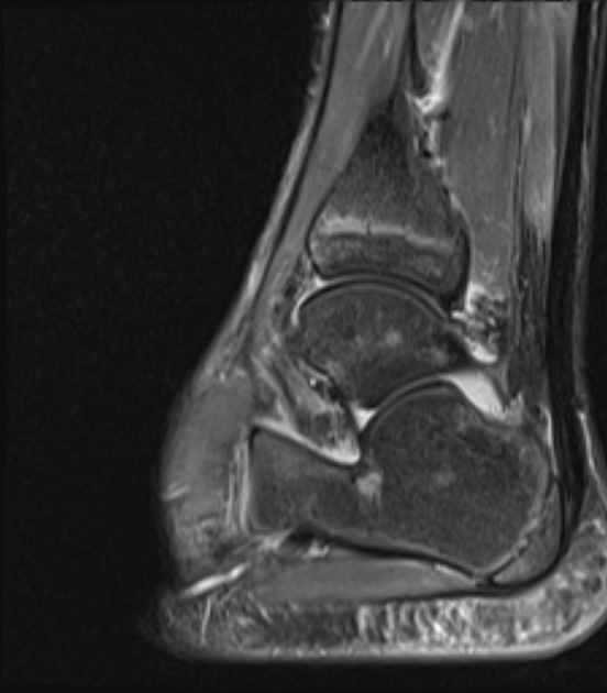
The spring (plantar calcaneonavicular) ligament complex is a group of ligaments that connect the calcaneum and navicular and support the talus.
On this page:
Gross anatomy
The spring ligament complex has three components:
-
superomedial ligament
- forms a sling, suspending/articulating against the head of the talus
- origin from anterior sustentaculum tali with a wide insertion onto the navicular
- merges with the inferior aspect of the tibiospring ligament (a portion of the superficial deltoid ligament), as best appreciated on coronal views 2
- strongest and most important longitudinal arch stabilizer; also most commonly torn/repaired
-
medioplantar oblique ligament
- also known as the lateral calcaneonavicular ligament
-
inferoplantar longitudinal ligament
- also known as the intermedial calcaneonavicular ligament
- minor role in stabilizing hindfoot and longitudinal arch 2
These ligaments act as the primary static stabilizers of the medial arch of the foot and, together with the posterior tibialis tendon (primary dynamic stabilizer), help support normal hindfoot relations.
Clinical importance
Failure of these stabilizers leads to hindfoot valgus and pes planus / pes planovalgus 2.
Related pathology
Quiz questions

References
- 1. Bloem HL et-al. Musculoskeletal Imaging, 2e (Expert Radiology). Saunders. ISBN:1455708135. Read it at Google Books - Find it at Amazon
- 2. Omar H, Saini V, Wadhwa V, Liu G, Chhabra A. Spring ligament complex: Illustrated normal anatomy and spectrum of pathologies on 3T MR imaging. (2016) European journal of radiology. 85 (11): 2133-2143. doi:10.1016/j.ejrad.2016.09.023 - Pubmed
Incoming Links
- Tibionavicular ligament
- Tibialis posterior muscle
- Longitudinal arch of the foot
- Spring ligament complex injury
- Adult acquired flatfoot disease
- Inferoplantar longitudinal ligament
- Midtarsal joint
- Medioplantar oblique ligament
- Talus
- Talonavicular joint
- Spring ligament recess
- Deltoid ligament of the ankle
- Sustentaculum tali
- MRI of the ankle (an approach)
- Superomedial calcaneonavicular ligament
- Tibiospring ligament
- Tibiocalcaneal ligament
Related articles: Anatomy: Lower limb
- skeleton of the lower limb
- joints of the lower limb
-
hip joint
- ligaments
- muscles
- additional structures
- hip joint capsule
- zona orbicularis
- iliotibial band
-
hip bursae
- anterior
- iliopsoas bursa (iliopectineal bursa)
- lateral
- subgluteal bursae
- greater trochanteric bursa (subgluteus maximus bursa)
- subgluteus medius bursa
- subgluteus minimus bursa
- gluteofemoral bursa
- subgluteal bursae
- postero-inferior
- anterior
- ossification centers
-
knee joint
- ligaments
- anterior cruciate ligament
- posterior cruciate ligament
- medial collateral ligament
- lateral collateral ligament
- meniscofemoral ligament (mnemonic)
-
posterolateral ligamentous complex
- arcuate ligament
- patellar tendon and quadriceps tendon
- anterolateral ligament
- posterior oblique ligament
- oblique popliteal ligament
- medial patellofemoral ligament
- additional structures
- extensor mechanism of the knee
- groove for the popliteus tendon
- knee bursae
- anterior bursae
- medial bursae
- lateral bursae
- posterior bursae
- knee capsule
- lateral patellar retinaculum
- medial patellar retinaculum
- menisci
- pes anserinus (mnemonic)
- ossification centers
- ligaments
- tibiofibular joints
-
ankle joint
- regional anatomy
- medial ankle
- lateral ankle
- anterior ankle
- ligaments
- medial collateral (deltoid) ligament
- lateral collateral ligament
- additional structures
- ankle bursae
- ossification centers of the ankle
- variants
- regional anatomy
- foot joints
- subtalar joint
- mid-tarsal (Chopart) joint
-
tarsometatarsal (Lisfranc) joint
- ligaments
- intermetatarsal joint
- metatarsophalangeal joint
- interphalangeal joint
- ossification centers
-
hip joint
- spaces of the lower limb
-
muscles of the lower limb
- muscles of the pelvic group
- muscles of the thigh
- muscles of the leg
- anterior compartment of the leg
- posterior compartments of the leg
- lateral compartment of the leg
- muscles of the foot
- dorsal muscles
- plantar muscles
- 1st layer
- 2nd layer
- 3rd layer
- 4th layer
- accessory muscles of the lower limb
- accessory gluteal muscles
-
accessory muscles of the ankle
- accessory peroneal muscles
- accessory flexor digitorum longus muscle
- accessory soleus muscle
- peroneocalcaneus internus muscle
- tibiocalcaneus internus muscle
- extensor hallucis capsularis tendon
- anterior fibulocalcaneus muscle
- accessory extensor digiti secundus muscle
- tibioastragalus anticus of Gruber muscle
- vascular supply of the lower limb
- arterial supply of the lower limb
- venous drainage of the lower limb
- innervation of the lower limb
- lymphatic system of the lower limb
- lymphatic pathways
- anteromedial group
- anterolateral group
- posteromedial group
- posterolateral group
- lower limb lymph nodes
- lymphatic pathways


 Unable to process the form. Check for errors and try again.