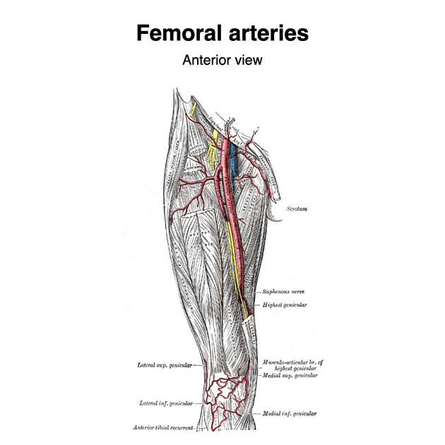
The femoral artery (FA) (TA: arteria femoralis) 6 is the continuation of the external iliac artery (EIA) below the level of the inguinal ligament. As well as supplying oxygenated blood to the lower limb, it gives off smaller branches to the anterior abdominal wall and superficial pelvis.
On this page:
Terminology
The femoral artery is commonly known clinically as the common femoral artery (CFA) and superficial femoral artery (SFA). The common femoral artery is the portion of the femoral artery between the inguinal ligament and branching of profunda femoris, and the superficial femoral artery is the portion distal to the branching of profunda femoris to the adductor hiatus.
The "superficial" adjective is considered by some as a misnomer as the artery itself runs deep in the mid-thigh, even though the nomenclature is logical in that it is to differentiate it from the profunda femoris artery which is even deeper 5! The Terminologia Anatomica and anatomists consider this division into common and superficial portions to be superfluous to requirements, and neither structure is given its own entry in the latest edition 6,7.
Summary
origin: continuation of the external iliac artery
course: courses posterior to the sartorius muscle and anterior to the femoral vein in the adductor canal exiting at the adductor hiatus to continue as the popliteal artery, giving off multiple muscular branches along its course
-
branches
superficial external pudendal artery
deep external pudendal artery
profunda femoris deep to adductor longus
-
relations
anterior: saphenous nerve, fibrous roof of adductor canal, vastus medialis, sartorius
posterior: femoral vein (upper part), adductor longus, profunda vessels, adductor magnus
lateral: femoral vein (lower part), vastus medialis, body of femur
-
supply
superficial pelvis
Gross anatomy
The femoral artery emerges underneath the inguinal ligament medial to the midpoint of the inguinal ligament and medial to the deep inguinal ring, halfway between the anterior superior iliac spine and symphysis pubis. The femoral vein lies medially.
The femoral artery runs down the front and medial side of the thigh with the first 4 cm of the vessel enclosed within the femoral sheath together with the femoral vein. Lateral but outside the sheath is the femoral nerve.
The femoral artery within the femoral triangle (~3-4 cm distal to the inguinal ligament) gives off the profunda femoris branch and ends as it passes through the adductor hiatus in adductor magnus to continue as the popliteal artery.
Relations
anterior: skin, superficial fascia, superficial iliac circumflex vein, superficial layer of fascia lata, anterior part of femoral sheath
posterior: posterior part of femoral sheath, pectineal fascia, psoas major tendon, capsule of hip joint, adductor longus, femoral vein (lower part of artery in femoral triangle)
lateral: femoral nerve
medial: femoral vein (upper part of artery)


 Unable to process the form. Check for errors and try again.