
Deep layer of the deep cervical fascia
Citation, DOI, disclosures and article data
At the time the article was created Francis Deng had no recorded disclosures.
View Francis Deng's current disclosuresAt the time the article was last revised Joachim Feger had no financial relationships to ineligible companies to disclose.
View Joachim Feger's current disclosures- Prevertebral fascia
- Perivertebral fascia
- Deep layer of deep cervical fascia (DLDCF)
- Prevertebral layer of deep cervical fascia
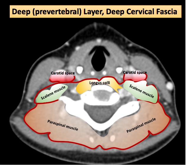
The deep layer of the deep cervical fascia is one of the three layers of the deep cervical fascia. It encases the paravertebral muscles and forms the perivertebral space. It consists of the perivertebral fascia (the anterior part of which is called the prevertebral fascia) and alar fascia 1-3.
Gross anatomy
Attachments
-
posteriorly
cervical vertebral spinous processes and transverse processes
-
laterally
first rib (from the portion of the layer called Sibson fascia)
-
superiorly
skull base
-
inferiorly
coccyx (for the prevertebral fascia)
visceral fascia between T2 and T4 (for the alar fascia)4
On each side, a flap attaches to the transverse processes of the cervical vertebrae and divides the peri-vertebral spaces into a pre-vertebral compartment anteriorly and a para-spinal compartment posteriorly 3.
Anteromedial to the scalene muscles, the deep layer splits into two leaves; the ventral leaf being the alar fascia, and the dorsal leaf being the prevertebral fascia (with the prevertebral space being between the prevertebral fascia and the spine). The space between the alar fascia and the prevertebral fascia is the danger space. The space between the alar fascia and the posterior aspect of the middle layer of the deep cervical fascia is the retropharyngeal space.
Contents
danger space (between prevertebral and alar fascia)
-
prevertebral space (anterior component of perivertebral space)
longus colli and capitis muscles
rectus capitis anterior and lateralis muscles
scalenus anterior, medius, and posterior muscles
sheath for subclavian artery and vein, brachial plexus
-
vertebral column
spinal cord and associated thecal sac, nerve roots, and vessels
vertebrae and associated discs and ligaments
-
paraspinal/paravertebral space (posterior component of perivertebral space)
deep cervical back muscles
-
pierced by the four cutaneous branches of the cervical plexus
In addition, all layers of the deep cervical fascia contribute to the carotid sheath.
References
- 1. Guidera A, Dawes P, Fong A, Stringer M. Head and Neck Fascia and Compartments: No Space for Spaces. Head Neck. 2014;36(7):1058-68. doi:10.1002/hed.23442 - Pubmed
- 2. Peter M. Som, Hugh D. Curtin. Head and Neck Imaging - 2 Volume Set. (2011) ISBN: 9780323053556 - Google Books
- 3. Kamalian S, Avery L, Lev M, Schaefer P, Curtin H, Kamalian S. Nontraumatic Head and Neck Emergencies. Radiographics. 2019;39(6):1808-23. doi:10.1148/rg.2019190159 - Pubmed
- 4. Gavid M, Dumollard J, Habougit C et al. Anatomical and Histological Study of the Deep Neck Fasciae: Does the Alar Fascia Exist? Surg Radiol Anat. 2018;40(8):917-22. doi:10.1007/s00276-018-1977-5 - Pubmed
Incoming Links
- Ascending cervical artery
- Alar fascia
- Ansa cervicalis
- Pharyngeal plexus
- Posterior cervical space
- Deep spaces of the head and neck
- Stellate ganglion block
- Perivertebral space
- Stellate ganglion
- Cervical plexus
- Supraclavicular triangle
- Supraclavicular nerves
- Carotid space
- Retropharyngeal space
- Visceral space
- Cloison sagittale
- Fascia
- Scalenus anterior muscle
- Middle cervical ganglion
- Deep cervical fascia
Related articles: Anatomy: Head and neck
- skeleton of the head and neck
-
cranial vault
- scalp (mnemonic)
- fontanelle
-
sutures
- calvarial
- facial
- frontozygomatic suture
- frontomaxillary suture
- frontolacrimal suture
- frontonasal suture
- temporozygomatic suture
- zygomaticomaxillary suture
- parietotemporal suture (parietomastoid suture)
- occipitotemporal suture (occipitomastoid suture)
- sphenofrontal suture
- sphenozygomatic suture
- spheno-occipital suture (not a true suture)
- lacrimomaxillary suture
- nasomaxillary suture
- internasal suture
- basal/internal
- skull landmarks
- frontal bone
- temporal bone
- parietal bone
- occipital bone
- skull base (foramina)
-
facial bones
- midline single bones
- paired bilateral bones
- cervical spine
- hyoid bone
- laryngeal cartilages
-
cranial vault
- muscles of the head and neck
- muscles of the tongue (mnemonic)
- muscles of mastication
-
facial muscles
- epicranius muscle
- circumorbital and palpebral muscles
- nasal muscles
-
buccolabial muscles
- elevators, retractors and evertors of the upper lip
- levator labii superioris alaeque nasalis muscle
- levator labii superioris muscle
- zygomaticus major muscle
- zygomaticus minor muscle
- levator anguli oris muscle
- malaris muscle
- risorius muscle
- depressors, retractors and evertors of the lower lip
- depressor labii inferioris muscle
- depressor anguli oris muscle
- mentalis muscle
- compound sphincter
-
orbicularis oris muscle
- incisivus labii superioris muscle
- incisivus labii inferioris muscle
-
orbicularis oris muscle
- muscle of mastication
- modiolus
- elevators, retractors and evertors of the upper lip
- muscles of the middle ear
- orbital muscles
- muscles of the soft palate
- pharyngeal muscles
- suprahyoid muscles
- infrahyoid muscles
- intrinsic muscles of the larynx
- muscles of the neck
- platysma muscle
- longus colli muscle
- longus capitis muscle
- scalenus anterior muscle
- scalenus medius muscle
- scalenus posterior muscle
- scalenus pleuralis muscle
- sternocleidomastoid muscle
-
suboccipital muscles
- rectus capitis posterior major muscle
- rectus capitis posterior minor muscle
- obliquus capitis superior muscle
- obliquus capitis inferior muscle
- accessory muscles of the neck
- deep cervical fascia
-
deep spaces of the neck
- anterior cervical space
- buccal space
- carotid space
- danger space
- deep cervical fascia
- infratemporal fossa
- masticator space
- parapharyngeal space
- stylomandibular tunnel
- parotid space
- pharyngeal (superficial) mucosal space
- perivertebral space
- posterior cervical space
- pterygopalatine fossa
- retropharyngeal space
- suprasternal space (of Burns)
- visceral space
- surgical triangles of the neck
- orbit
- ear
- paranasal sinuses
- upper respiratory tract
- viscera of the neck
- blood supply of the head and neck
-
arterial supply
-
common carotid artery
- carotid body
- carotid bifurcation
- subclavian artery
- variants
-
common carotid artery
- venous drainage
-
arterial supply
- innervation of the head and neck
-
cranial nerves
- olfactory nerve (CN I)
- optic nerve (CN II)
- oculomotor nerve (CN III)
- trochlear nerve (CN IV)
-
trigeminal nerve (CN V) (mnemonic)
- trigeminal ganglion
- ophthalmic division
- maxillary division
- mandibular division
- abducens nerve (CN VI)
- facial nerve (CN VII)
-
vestibulocochlear nerve (CN VIII)
- vestibular ganglion (Scarpa's ganglion)
- glossopharyngeal nerve (CN IX)
- vagus nerve (CN X)
- (spinal) accessory nerve (CN XI)
- hypoglossal nerve (CN XII)
- parasympathetic ganglia of the head and neck
- cervical sympathetic ganglia
- greater occipital nerve
- third occipital nerve
-
cervical plexus
- muscular branches
- longus capitis
- longus colli
- scalenes
- geniohyoid
- thyrohyoid
-
ansa cervicalis
- omohyoid (superior and inferior bellies separately)
- sternothyroid
- sternohyoid
- phrenic nerve
- contribution to the accessory nerve (CN XI)
- cutaneous branches
- muscular branches
- brachial plexus
- pharyngeal plexus
-
cranial nerves
- lymphatic drainage of the head and neck
- embryological development of the head and neck


 Unable to process the form. Check for errors and try again.