
The hamate is one of the carpal bones, forms part of the distal carpal row and has a characteristic hook on its volar surface.
On this page:
Gross anatomy
Osteology
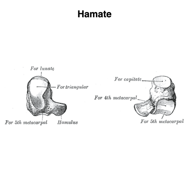
The hamate has a wedge-shaped body. It bears an uncinate (unciform) hamulus (hook of hamate) which projects in a volar fashion from the distal part of its palmar surface. It is the most ulnar based bone within the distal carpal row. It has four surfaces:
superior surface: apex of the wedge is narrow, convex, and smooth
inferior surface: concave facets separated by a ridge
volar surface: hook-like process (hamulus) on the lower and ulnar side
medial surface: oblong facet
Articulations
The hamate articulates with five bones: the lunate, capitate, triquetral, fourth metacarpal, and fifth metacarpal:
proximal thin margin of the wedge articulates with the lunate while the fourth and fifth metacarpals articulate with the base distally
medial surface is broad and articulates with the triquetrum
lateral surface articulates with the capitate
hook of the hamate is distal and radial in relation to the pisiform
Attachments
Musculotendinous
flexor digiti minimi brevis and opponens digiti minimi both arise from the convex surface of the hook of the hamate
abductor digiti minimi arises from the hook of hamate
flexor carpi ulnaris inserts via the pisohamate ligament into apex of the hook of hamate
flexor digitorum profundus uses the hook of hamate as a pulley during flexion of the 5th finger with ulnar deviation
Ligamentous
flexor retinaculum attaches to the apex of the hamulus
pisohamate ligament
triquetrohamate ligament
capitohamate ligament
Arterial supply
Nearly 100% of hamate specimens have radial artery supply. Most specimens have dual radial and ulnar supply with the ulnar artery contributing a smaller percentage. ~30% have absent ulnar artery supply.
Innervation
A terminal deep branch of the ulnar nerve lies in a transverse groove underneath the hook of the hamate towards its base.
Variant anatomy
os hamuli proprium (incomplete fusion of the ossification center of the hook)
capitohamate (carpal coalition between the hamate and capitate)
Development
Ossification
It is the second bone to ossify following the capitate. It is thought to start ossifying by the end of the third month. Fusion is complete by age 14-16.
History and etymology
The term derives from the Latin hamatus meaning hooked.
Related investigations
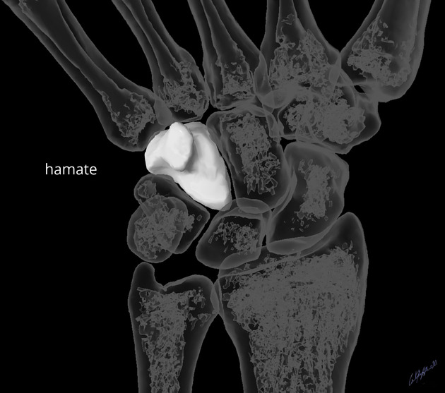
Plain radiograph
The hamate may be visualized on a number of series of the distal upper limb including:
Routine PA and lateral views of the wrist potentially miss hamate fractures. Carpal tunnel views, in addition to PA, lateral, and oblique views are typically required when hamate fractures are suspected.
Cross-sectional imaging
CT has a sensitivity of 100% and a specificity of 94% in visualizing fractures of the hamate compared to x-ray which has a sensitivity of 72% and specificity of 88%.4


 Unable to process the form. Check for errors and try again.