Subacromial-subdeltoid bursa
Citation, DOI, disclosures and article data
At the time the article was created Jeremy Jones had no recorded disclosures.
View Jeremy Jones's current disclosuresAt the time the article was last revised Joshua Yap had no financial relationships to ineligible companies to disclose.
View Joshua Yap's current disclosures- SA-SD bursa
- SA-SDB
- Subacromial bursa
- SASD bursa
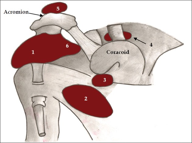
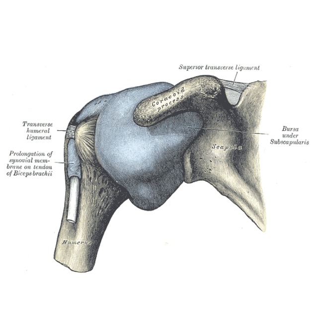
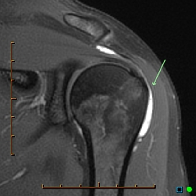
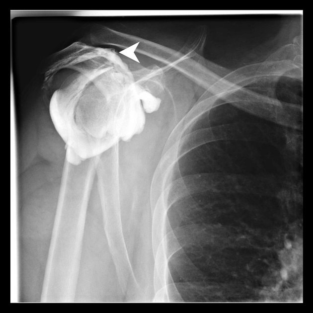
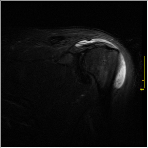
The subacromial-subdeltoid bursa (SASD), also simply known as the subacromial bursa, is a bursa within the shoulder that is simply a potential space in normal individuals.
On this page:
Gross anatomy
-
found deep to the deltoid muscle and the coraco-acromial arch
medially in close relation to the acromio-clavicular joint
the mid-point of the bursa commonly corresponds to the most anterior extent of the acromion
-
extends laterally over the greater tubercle of the humerus, terminating in its sub-deltoid reflection an average of 4 cm from the mid-point of the acromion
this osseous landmark also corresponds to the posterior bursal reflection
envelops the bicipital groove as it extends inferiorly
-
superficial anatomic relations include the rotator interval and the supraspinatus tendon
invested by parallel layers of peri-bursal fat
relationship with supraspinatus tendon consistent, whereas it may or may not interface with the infraspinatus
-
anatomically discontinuous with the gleno-humeral joint
communication may occur in the presence of rotator cuff pathology
ADVERTISEMENT: Supporters see fewer/no ads
Radiographic features
Ultrasound
-
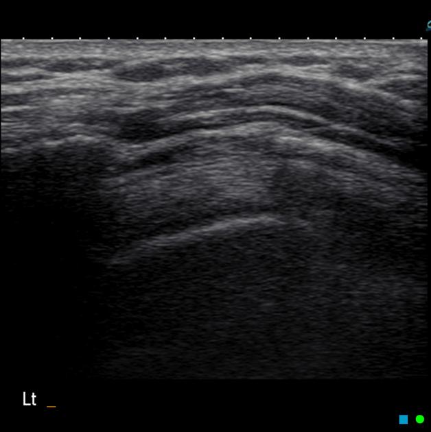
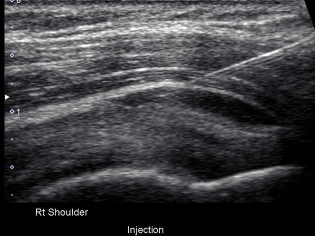
the hyperechoic peri-bursal fat forms a prominent interface between the deltoid and/or acromion and the underlying supraspinatus allowing identification of the potential space representing the bursa
anechoic fluid may interpose between the layers of peri-bursal fat forming a tri-laminar interface
bursal thickness is typically less than 1 mm in the presence of physiologic amounts of bursal fluid
color flow Doppler examination should reveal an absence of bursal vascularity
dynamic visualization during shoulder abduction and adduction should demonstrate smooth translocation of the bursal reflection beneath the acromion
Related pathology
-
bursitis
septic bursitis
rheumatoid arthritis
Quiz questions
References
- 1. Hirji Z, Hunjun JS, Choudur HN. Imaging of the bursae. J Clin Imaging Sci. 2011;1 (1): 22. doi:10.4103/2156-7514.80374 - Free text at pubmed - Pubmed citation
- 2. Beals T, Harryman D, Lazarus M. Useful Boundaries of the Subacromial Bursa. Arthroscopy. 1998;14(5):465-70. doi:10.1016/s0749-8063(98)70073-8 - Pubmed
- 3. Messina C, Banfi G, Orlandi D et al. Ultrasound-Guided Interventional Procedures Around the Shoulder. BJR. 2016;89(1057):20150372. doi:10.1259/bjr.20150372 - Pubmed
- 4. Pourcho A, Colio S, Hall M. Ultrasound-Guided Interventional Procedures About the Shoulder. Phys Med Rehabil Clin N Am. 2016;27(3):555-72. doi:10.1016/j.pmr.2016.04.001 - Pubmed
Incoming Links
- Calcific tendinitis
- Bursa
- Shoulder injury related to vaccine administration
- Ultrasound of the shoulder
- Shoulder
- Coracoacromial ligament
- Subacromial impingement
- Shoulder bursae
- Medical abbreviations and acronyms (S)
- Fosbury flop tear of the rotator cuff
- Subacromial-subdeltoid bursitis
- Acromion
- Glenohumeral joint
- Full-thickness rotator cuff tear
- Acromioclavicular joint cyst
- Partial-thickness rotator cuff tear
- Coracoacromial arch
- Subcoracoid bursa
- Bursal-sided rotator cuff tear
- Adhesive capsulitis of the shoulder
- Migration of supraspinatus tendon calcification
- Hydroxyapatite deposition disease
- Subacromial-subdeltoid bursal rupture
- Proximal humerus fracture
- Shoulder bursa (Gray's illustration)
- Anterior labroligamentous periosteal sleeve avulsion lesion (ALPSA)
- Iatrogenic injury of the spinal accessory nerve
- Subscapularis tendon tear
- Acromioclavicular joint injury with muscle injuries (ultrasound)
- Suprascapular neuropathy
- Full-thickness rotator cuff tear
- Subacromial subdeltoid bursa injection (ultrasound-guided)
- Subacromial-subdeltoid bursitis with rotator cuff tears
- Bifid biceps tendon
- Partial supraspinatus tear wih subacromial impingement
- Subacromial impingement
- Bony Bankart injury with Hill-Sachs
- Bifid biceps tendon
- Impingement syndrome with supraspinatus tendinopathy and atrophy
Related articles: Anatomy: Upper limb
-
skeleton of the upper limb[+][+]
- clavicle
- scapula
- humerus
- radius
- ulna
- hand
- accessory ossicles of the upper limb
- accessory ossicles of the shoulder
- accessory ossicles of the elbow
-
accessory ossicles of the wrist (mnemonic)
- os centrale carpi
- os epilunate
- os epitriquetrum
- os styloideum
- os hamuli proprium
- lunula
- os triangulare
- trapezium secondarium
- os paratrapezium
- os radiostyloideum (persistent radial styloid)
- joints of the upper limb
-
pectoral girdle
-
shoulder joint
- articulations[+][+]
- associated structures
- joint capsule[+][+]
-
bursae
- subacromial-subdeltoid (SASD) bursa
- subscapular recess
- subcoracoid bursa
- coracoclavicular bursa
- supra-acromial bursa
- ligaments[+][+]
- movements[+][+]
- scapulothoracic joint
-
glenohumeral joint
- arm flexion
- arm extension
- arm abduction
- arm adduction
- arm internal rotation (medial rotation)
- arm external rotation (lateral rotation)
- circumduction
- arterial supply - scapular anastomosis
- ossification centers
-
shoulder joint
-
elbow joint[+][+]
- proximal radioulnar joint
- ligaments
- associated structures
- movements
- alignment
- arterial supply - elbow anastomosis
- development
-
wrist joint[+][+]
- articulations
-
ligaments
- intrinsic ligaments
- extrinsic ligaments
- radioscaphoid ligament
- dorsal intercarpal ligament
- dorsal radiotriquetral ligament
- dorsal radioulnar ligament
- volar radioulnar ligament
- radioscaphocapitate ligament
- long radiolunate ligament
- Vickers ligament
- short radiolunate ligament
- ulnolunate ligament
- ulnotriquetral ligament
- ulnocapitate ligament
- ulnar collateral ligament
- associated structures
- extensor retinaculum
- flexor retinaculum
- joint capsule
- movements
- alignment
- ossification centers
-
hand joints[+][+]
- articulations
- carpometacarpal joint
-
metacarpophalangeal joints
- palmar ligament (plate)
- collateral ligament
-
interphalangeal joints
- palmar ligament (plate)
- collateral ligament
- movements
- ossification centers
- articulations
-
pectoral girdle
- spaces of the upper limb[+][+]
- muscles of the upper limb[+][+]
- shoulder girdle
- anterior compartment of the arm
- posterior compartment of the arm
-
anterior compartment of the forearm
- superficial
- intermediate
- deep
-
posterior compartment of the forearm (extensors)
- superficial
- deep
- muscles of the hand
-
accessory muscles
- elbow
- volar wrist midline
- palmaris longus profundus
- aberrant palmaris longus
- volar wrist radial-side
- accessory flexor digitorum superficialis indicis
- flexor indicis profundus
- flexor carpi radialis vel profundus
- accessory head of the flexor pollicis longus (Gantzer muscle, common)
- volar wrist ulnar-side
- dorsal wrist
- blood supply to the upper limb[+][+]
-
arteries
- subclavian artery (mnemonic)
- axillary artery
- brachial artery (proximal portion)
- ulnar artery
- radial artery
- veins
-
arteries
- innervation of the upper limb[+][+]
- intercostobrachial nerve
-
brachial plexus (mnemonic)
- branches from the roots
- branches from the trunks
- branches from the cords
- lateral cord
- posterior cord
- medial cord
- terminal branches
- lymphatic drainage of the upper limb[+][+]




 Unable to process the form. Check for errors and try again.