
The inferior vena cava (IVC) (plural: inferior venae cavae) is one of the great vessels that drains venous blood from the lower limbs, pelvis and abdomen into the right atrium of the heart.
On this page:
Gross anatomy
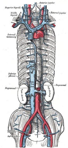
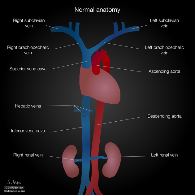
The inferior vena cava is formed by the confluence of the two common iliac veins at the L5 vertebral level. The IVC has a retroperitoneal course within the abdominal cavity. It runs along the right side of the vertebral column with the aorta lying laterally on the left. Various other veins drain into the IVC along its course before it passes through the diaphragm at the caval hiatus at the T8 level. Its intrathoracic course is very short before draining into the right atrium at the inferior cavoatrial junction.
Tributaries
T8: paired inferior phrenic veins
T8: hepatic veins 3
L1: right suprarenal vein
L1: renal veins
L2: right gonadal vein
L1-L5: lumbar veins
L5: common iliac veins (origin)
Since the IVC is not a midline structure, there is a degree of asymmetry of drainage, e.g. the gonadal and suprarenal veins drain into the IVC on the right side, but into the left renal vein on the left.
Relations
anterior: right common iliac artery, right gonadal vessels, third part of duodenum (D3), common bile duct, portal vein, head of pancreas, first part of duodenum (D1), epiploic foramen, liver
posterior: lumbar vertebrae, intervertebral discs, anterior longitudinal ligament, right psoas major, right sympathetic trunk, celiac plexus, right lumbar arteries, right renal artery, right suprarenal arteries, right inferior phrenic artery
lateral (left): abdominal aorta, caudate lobe of the liver, right crus
lateral (right): right kidney, right ureter, second part of duodenum (D2), liver
Development
The normal IVC has a complex embryological development with many embryological veins contributing to many different parts:
right vitelline vein: forms suprahepatic and hepatic segments of IVC
right subcardinal vein: forms suprarenal segment
right subsupracardinal anastomosis: forms renal segment
right supracardinal vein: forms infrarenal segment
right posterior cardinal vein: forms distal most IVC and its bifurcation into common iliac veins
Variant anatomy
Inferior caval abnormalities are typically the result of abnormal embryologic development involving the vitelline, posterior cardinal, subcardinal and supracardinal veins 3:
absence of IVC (entire or only the infrarenal segment)
azygos continuation of the IVC
extrahepatic portocaval shunt (Abernethy malformation)
Rarely a Eustachian valve at the inferior cavoatrial junction may be present.


 Unable to process the form. Check for errors and try again.