Navicular
Citation, DOI, disclosures and article data
At the time the article was created Laura Smith had no recorded disclosures.
View Laura Smith's current disclosuresAt the time the article was last revised Calum Worsley had no financial relationships to ineligible companies to disclose.
View Calum Worsley's current disclosures- Navicular bone
- Tarsal navicular
- Navicular bones
- Tarsal scaphoid
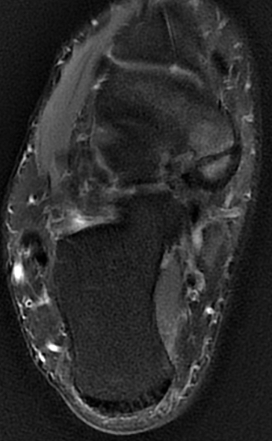
The navicular bone is found in the midfoot and is one of the tarsal bones. Its structure resembles that of a boat. It is the last bone of the foot to ossify fully 1.
On this page:
Summary
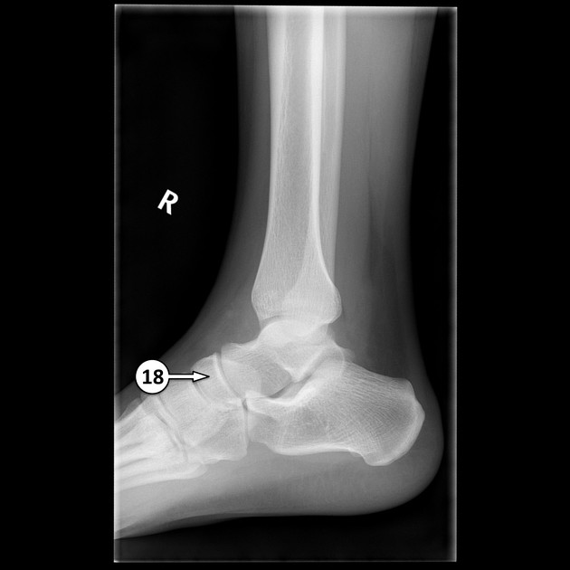
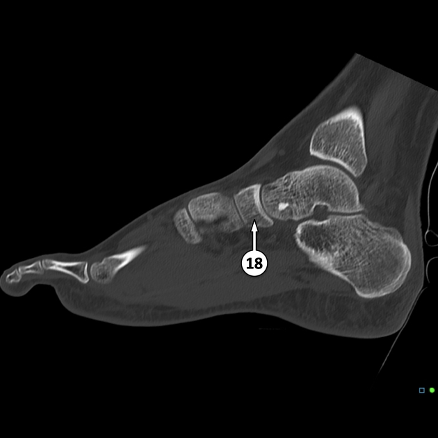
location: lies within the medial aspect of the midfoot
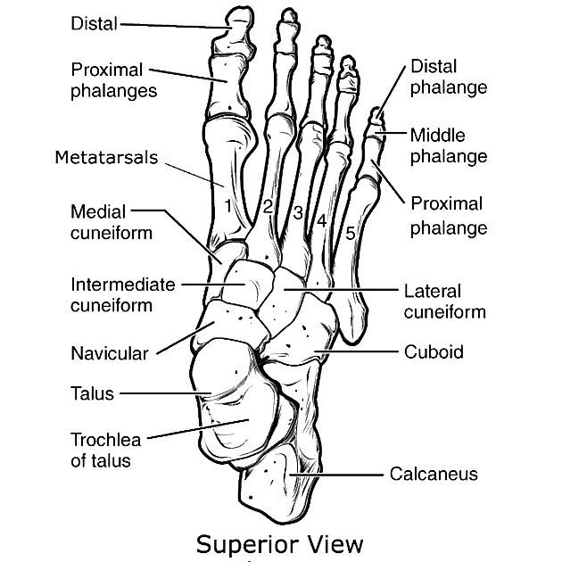
relations: the talus bone, cuboid bone and the three cuneiform bones
arterial supply: branches of the dorsalis pedis, posterior tibial and medial plantar arteries 4
Gross anatomy
Articulations
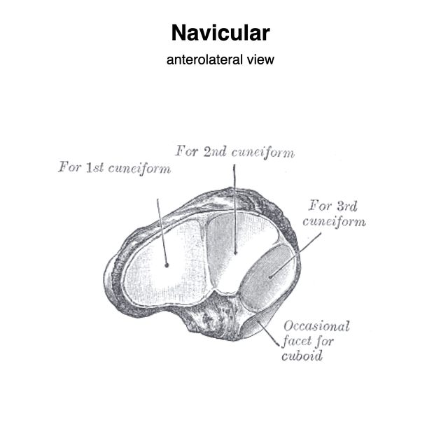
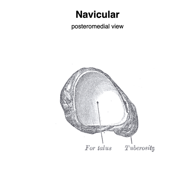
The navicular bone articulates proximally with the head of the talus bone, laterally with the cuboid bone and also distally to the lateral, intermediate and medial cuneiform bones 2.
Attachments
It gives attachments to the superomedial and inferior calcaneonavicular ligament which together form the spring ligament within the foot 2.
Musculotendinous
The tibialis posterior tendon inserts into the medial side of the navicular bone 2.
Ligamentous
The dorsal cuneonavicular ligament and plantar cuneonavicular ligament are two ligaments that join each cuneiform bone to the navicular. The medial cuneiform bone also is joined to the navicular by the medial cuneonavicular ligament 3.
ADVERTISEMENT: Supporters see fewer/no ads
Arterial supply
A branch of dorsalis pedis artery gives off three to five smaller branches which supply the navicular from the medial side. On the lateral aspect of the bone, small branches arise from the posterior tibial artery. Branches of the medial plantar artery also supply the plantar surface of the bone 4. In the central navicular there is a region of watershed blood supply which predisposes the bone to stress fractures.
Variant anatomy
An accessory ossicle can be present on the medial aspect of the navicular known as an accessory navicular.
Development
Abnormalities of the ossification of the navicular are common. The navicular is the last bone in the foot to ossify. The navicular anlage ossifies in girls between 18-24 months, and in boys between 30-36 months 4.
Related pathology
Quiz questions
References
- 1. Stuart L. Weinstein, Joseph A. Buckwalter. Turek's Orthopaedics. (2005) ISBN: 9780781742986 - Google Books
- 2. Golano P, Fariñas O, Sáenz I. The Anatomy of the Navicular and Periarticular Structures. Foot Ankle Clin. 2004;9(1):1-23. doi:10.1016/S1083-7515(03)00155-4 - Pubmed
- 3. Armen S. Kelikian, Shahan K. Sarrafian. Sarrafian's Anatomy of the Foot and Ankle. (2011) ISBN: 9780781797504 - Google Books
- 4. Trueta J. Studies of the Development and Decay of the Human Frame. Butterworth-Heinemann; 2014. ISBN: 978-1-4831-6802-9.
Incoming Links
- Chopart fracture-dislocation
- Midfoot
- Medioplantar oblique ligament
- Subtalar dislocation
- Bifurcate ligament
- Talonavicular joint
- Müller-Weiss syndrome
- Tibioastragalus anticus of Gruber muscle
- Flat-top talus
- Cuneonavicular joint
- Alban Köhler
- Cornuate navicular
- Stress response
- Cuboid
- Fatigue fracture
- Foot (dorsoplantar view)
- Midtarsal sprain
- Tibialis posterior muscle
- Congenital talipes equinovarus
- Mueller-Weiss syndrome (classification)
- Accessory navicular
- Kohler disease
- Navicular osteonecrosis - paediatric
- Tarsal navicular fracture
- Navicular fracture
- Cuboid-navicular coalition
- Talonavicular coalition
- Charcot neuroarthropathy of the foot
- Neglected congenital talipes equinovarus
- Navicular fracture
- Absence of tarsal navicular bone - bilateral
- Accessory navicular bone - type II
- Navicular (Gray's illustration)
- Dorsal avulsion fracture of the navicular bone
- Midtarsal sprain
- Foot avulsion injuries
- Köhler disease
- Mueller Weiss syndrome
- Dorsal avulsion fracture of navicular bone
- Foot radiograph (anatomy quiz)
Related articles: Anatomy: Lower limb
- skeleton of the lower limb
- joints of the lower limb
-
hip joint
- ligaments
- muscles
- additional structures
- hip joint capsule
- zona orbicularis
- iliotibial band
-
hip bursae
- anterior
- iliopsoas bursa (iliopectineal bursa)
- lateral
- subgluteal bursae
- greater trochanteric bursa (subgluteus maximus bursa)
- subgluteus medius bursa
- subgluteus minimus bursa
- gluteofemoral bursa
- subgluteal bursae
- postero-inferior
- anterior
- ossification centers
-
knee joint
- ligaments
- anterior cruciate ligament
- posterior cruciate ligament
- medial collateral ligament
- lateral collateral ligament
- meniscofemoral ligament (mnemonic)
-
posterolateral ligamentous complex
- arcuate ligament
- patellar tendon and quadriceps tendon
- anterolateral ligament
- posterior oblique ligament
- oblique popliteal ligament
- medial patellofemoral ligament
- additional structures
- extensor mechanism of the knee
- groove for the popliteus tendon
- knee bursae
- anterior bursae
- medial bursae
- lateral bursae
- posterior bursae
- knee capsule
- lateral patellar retinaculum
- medial patellar retinaculum
- menisci
- pes anserinus (mnemonic)
- ossification centers
- ligaments
- tibiofibular joints
-
ankle joint
- regional anatomy
- medial ankle
- lateral ankle
- anterior ankle
- ligaments
- medial collateral (deltoid) ligament
- lateral collateral ligament
- additional structures
- ankle bursae
- ossification centers of the ankle
- variants
- regional anatomy
- foot joints
- subtalar joint
- mid-tarsal (Chopart) joint
-
tarsometatarsal (Lisfranc) joint
- ligaments
- intermetatarsal joint
- metatarsophalangeal joint
- interphalangeal joint
- ossification centers
-
hip joint
- spaces of the lower limb
-
muscles of the lower limb
- muscles of the pelvic group
- muscles of the thigh
- muscles of the leg
- anterior compartment of the leg
- posterior compartments of the leg
- lateral compartment of the leg
- muscles of the foot
- dorsal muscles
- plantar muscles
- 1st layer
- 2nd layer
- 3rd layer
- 4th layer
- accessory muscles of the lower limb
- accessory gluteal muscles
-
accessory muscles of the ankle
- accessory peroneal muscles
- accessory flexor digitorum longus muscle
- accessory soleus muscle
- peroneocalcaneus internus muscle
- tibiocalcaneus internus muscle
- extensor hallucis capsularis tendon
- anterior fibulocalcaneus muscle
- accessory extensor digiti secundus muscle
- tibioastragalus anticus of Gruber muscle
- vascular supply of the lower limb
- arterial supply of the lower limb
- venous drainage of the lower limb
- innervation of the lower limb
- lymphatic system of the lower limb
- lymphatic pathways
- anteromedial group
- anterolateral group
- posteromedial group
- posterolateral group
- lower limb lymph nodes
- lymphatic pathways