
The parapharyngeal space (PPS), also known as the prestyloid parapharyngeal space, is a deep compartment of the head and neck around which most other suprahyoid fascial spaces are arranged. It consists largely of fat, neurovascular structures, and, in some definitions, the retromandibular part of the deep lobe of the parotid gland.
On this page:
Terminology
Two naming conventions exist in the literature. In the first definition, familiar to most head and neck surgeons, the parapharyngeal space is divided into prestyloid and poststyloid (retrostyloid) compartments 1-3,10. In the second definition, introduced by some radiologists, the prestyloid parapharygeal space is simply termed the parapharyngeal space, and the poststyloid pharapharygeal space is termed the carotid space 4-6. The latter facilitates differential diagnosis and is used in this article.
Other terms for the parapharyngeal space include the lateral pharyngeal space, pharyngomaxillary space, and even less commonly pterygomaxillary space, pterygopharyngeal space, peripharyngeal space, and pharyngomasticatory space 1. The term pterygomandibular space has rarely been used for this location, but this term is best reserved for the subcompartment of the masticator space instead.
Gross anatomy
The parapharyngeal space is shaped like an inverted pyramid, with its base at the skull base, with its apex inferiorly pointing towards the greater cornu of the hyoid bone 2.
Contents
fat (main component)
-
vessels
internal maxillary artery, depending on its course
ascending pharyngeal artery, depending on its course
pterygoid venous plexus, in its portion surrounded by fat (the rest is within the masticator space)
-
nerve
small branch of the mandibular division of the trigeminal nerve (cranial nerve V3) supplying the tensor veli palatini muscle 15
-
salivary tissue
minor or ectopic salivary gland/rests
retromandibular portion of the deep lobe of parotid gland, in some definitions 1-3
Lymph nodes and muscle are not included in the radiological definition of the parapharyngeal space.
A mnemonic to remember the contents of the parapharyngeal space is FATPIG.
Boundaries
The parapharyngeal space has complex fascial margins occupying the space between the muscles of mastication and the muscles of deglutition 1-6:
superior: base of skull
inferior: greater cornu of the hyoid bone, although some state the space functionally ends higher, with the styloglossus muscle at the level of the angle of the mandible 1
medial: middle layer of the deep cervical fascia covering the superior pharyngeal constrictor and levator and tensor veli palatini muscles
lateral: superficial layer of the deep cervical fascia extending between styloid process and mandibular ramus, covering the parotid and lateral pterygoid muscle, although some state the fascia creates the stylomandibular tunnel through which part of the deep lobe of the parotid protrudes into the parapharyngeal space 1
anterior: pterygomandibular raphe and superficial layer of the deep cervical fascia covering the medial pterygoid muscle
posterior: an extension of tensor veli palatini muscle fascia termed the tensor-vascular-styloid fascia 1,10-12; or an extension of the fascia of the stylopharyngeus, styloglossus, and levator veli palatini muscles 13,14
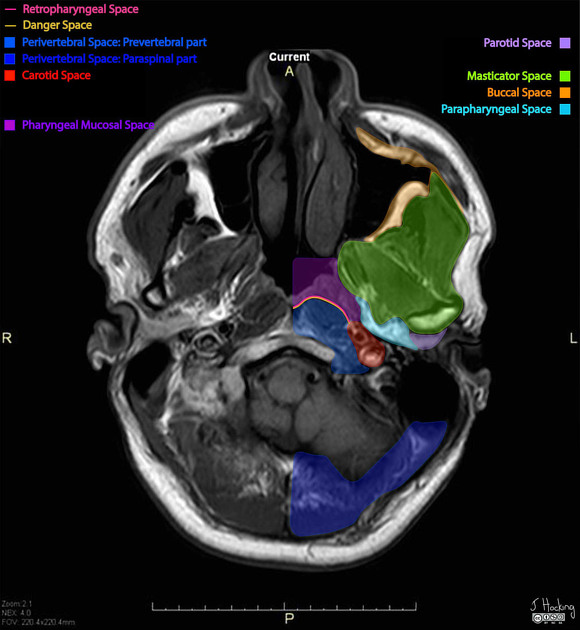
Relations
posteromedial to the masticator space, particularly medial pterygoid muscle
anteromedial to the parotid space
posterolateral to the pharyngeal mucosal space
anterolateral to the prevertebral space, retropharyngeal space, and danger space
anterior to the carotid (poststyloid parapharyngeal) space
overlaps with the infratemporal fossa 5
Radiographic features
CT/MRI
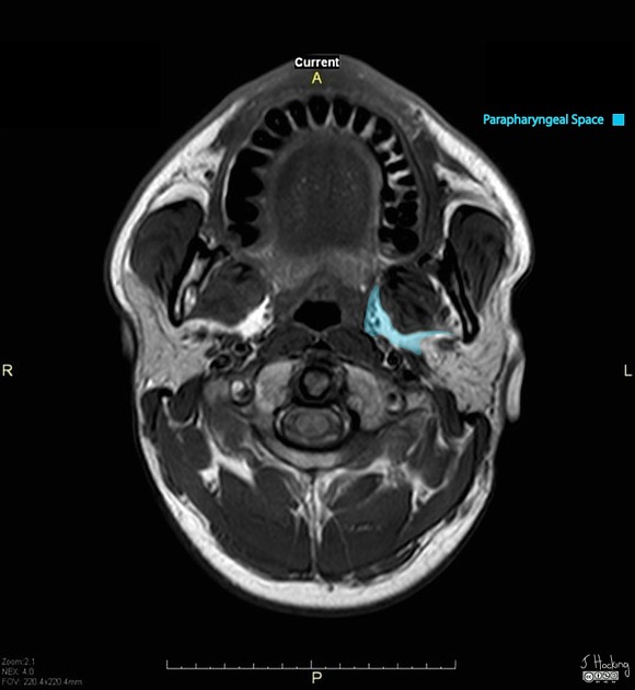
The parapharyngeal space appears triangular in the axial plane with density/signal consistent with fat.
Knowledge about the displacement patterns of fat within the parapharyngeal space will aid in the localization of lesions within adjacent deep spaces of the head and neck. A lesion arising in the:
masticator space displaces the parapharyngeal fat posteromedially
parotid space displaces the parapharyngeal fat anteromedially
pharyngeal mucosal space displaces the parapharyngeal fat posterolaterally
retropharyngeal space, danger space, and prevertebral space displace the parapharyngeal fat anterolaterally
carotid (poststyloid parapharyngeal) space displaces the parapharyngeal fat anteriorly
In contrast, a lesion primarily involving the parapharyngeal space will displace the carotid space posteriorly and the pharynx medially.
Related pathology
Lesions involving the (prestyloid) parapharyngeal space include 7:
-
tumors 8,9
salivary gland tumors (most common), most commonly pleomorphic adenoma
neurogenic tumors, such as trigeminal schwannoma
parapharyngeal abscess or phlegmon/cellulitis
-
developmental lesions
second branchial cleft cyst (atypical location)


 Unable to process the form. Check for errors and try again.